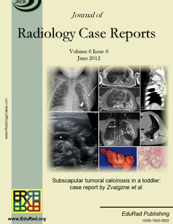
Subscapular tumoral calcinosis in a toddler: case report
DOI:
https://doi.org/10.3941/jrcr.v6i6.967Keywords:
Tumoral Calcinosis, Magnetic Resonance Imaging, Computed Tomography, Case ReportAbstract
Tumoral calcinosis is uncommon in toddlers, and rare within the subscapular area. Although typically benign, tumoral calcinosis is often incorrectly diagnosed prior to biopsy. We present a case of subscapular tumoral calcinosis in a 16-month old girl and discuss the radiological findings on X-ray, ultrasound, computed tomography and magnetic resonance imaging, including the first description of T1-weighted post contrast imaging, which demonstrate the fibrotic components of tumoral calcinosis.Downloads
Published
2012-05-23
Issue
Section
Pediatric Radiology
License
The publisher holds the copyright to the published articles and contents. However, the articles in this journal are open-access articles distributed under the terms of the Creative Commons Attribution-NonCommercial-NoDerivs 4.0 License, which permits reproduction and distribution, provided the original work is properly cited. The publisher and author have the right to use the text, images and other multimedia contents from the submitted work for further usage in affiliated programs. Commercial use and derivative works are not permitted, unless explicitly allowed by the publisher.


