Magnetic Resonance Angiography as a Radiation-Free Alternative for Pulmonary Embolism: A Longitudinal Case Report
DOI:
https://doi.org/10.3941/jrcr.6169Abstract
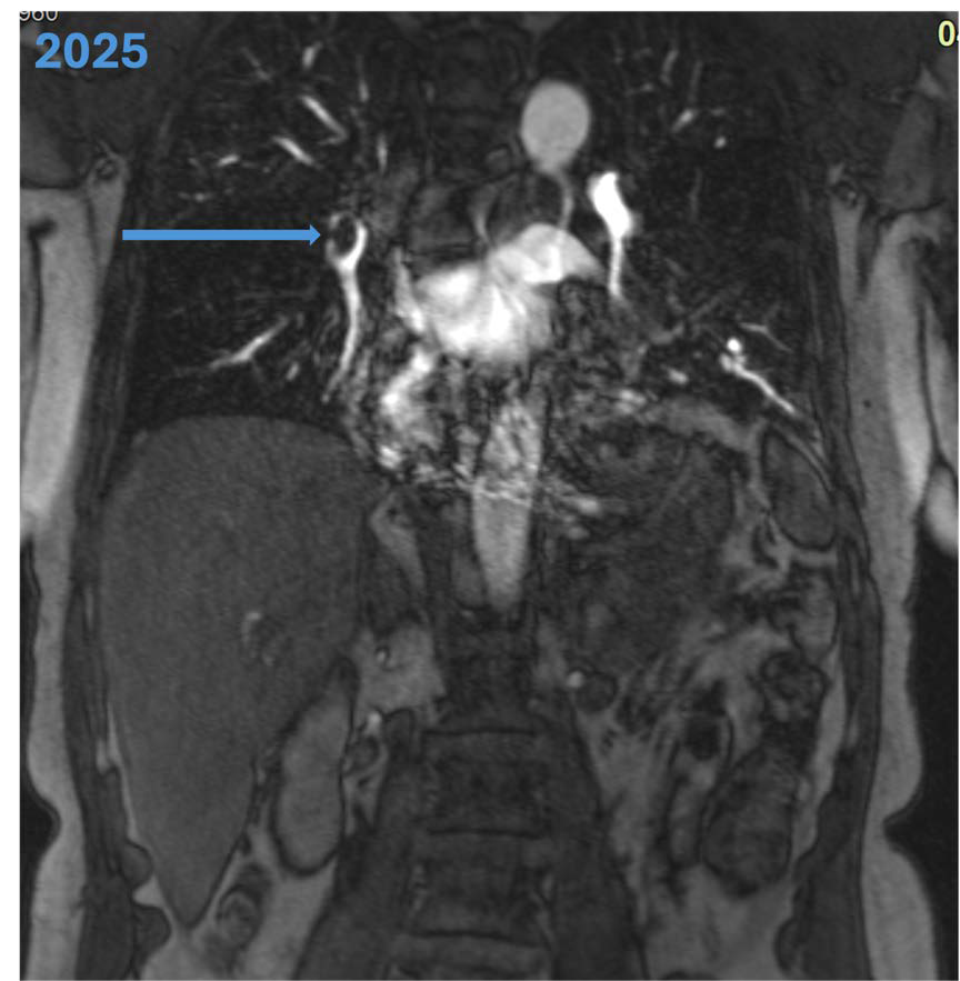
Pulmonary embolism is life-threatening and is usually diagnosed with computed tomography (CT), which involves ionizing radiation and iodinated contrast. This case describes a pulmonary embolism diagnosed with magnetic resonance angiography (MRA) in 2004 and 2025, providing a longitudinal comparison of MRA/MRI’s reproducibility in pulmonary embolism and venous thromboembolism diagnosis in this patient.
A 65-year-old female with prior pulmonary embolism and chronic venous insufficiency presented with dyspnea, pleuritic chest pain and leg edema. Due to patient concerns about radiation from CT angiography, MRI was employed first line.MRI revealed multiple pulmonary emboli, lower limb deep vein thromboses and infarction pneumonia. The patient was treated with anticoagulation and antibiotics.
This case shows MRA can diagnose pulmonary embolism while avoiding radiation. It underscores MRI’s role as a safe diagnostic alternative to CT and supports its uptake for the broader population.
Downloads
Published
Issue
Section
License
Copyright (c) 2026 Journal of Radiology Case Reports

This work is licensed under a Creative Commons Attribution-NonCommercial-NoDerivatives 4.0 International License.
The publisher holds the copyright to the published articles and contents. However, the articles in this journal are open-access articles distributed under the terms of the Creative Commons Attribution-NonCommercial-NoDerivs 4.0 License, which permits reproduction and distribution, provided the original work is properly cited. The publisher and author have the right to use the text, images and other multimedia contents from the submitted work for further usage in affiliated programs. Commercial use and derivative works are not permitted, unless explicitly allowed by the publisher.