
Ga-68 DOTATATE PET/CT in a 19-Year-Old Male with Left Optic Nerve Sheath Meningioma: Diagnostic Utility in Differentiating Inflammatory Perineuritis
DOI:
https://doi.org/10.3941/jrcr.6102Abstract
Background: Optic nerve sheath meningioma is an uncommon tumour arising from the meningothelial cells of the optic nerve sheath. Diagnostic challenges can arise when differentiating optic nerve sheath meningioma and optic neuritis due to similar clinical presentation and magnetic resonance imaging findings. A delay in diagnosis can lead to a delay in appropriate therapy thereby increasing the risk of irreversible vision loss.
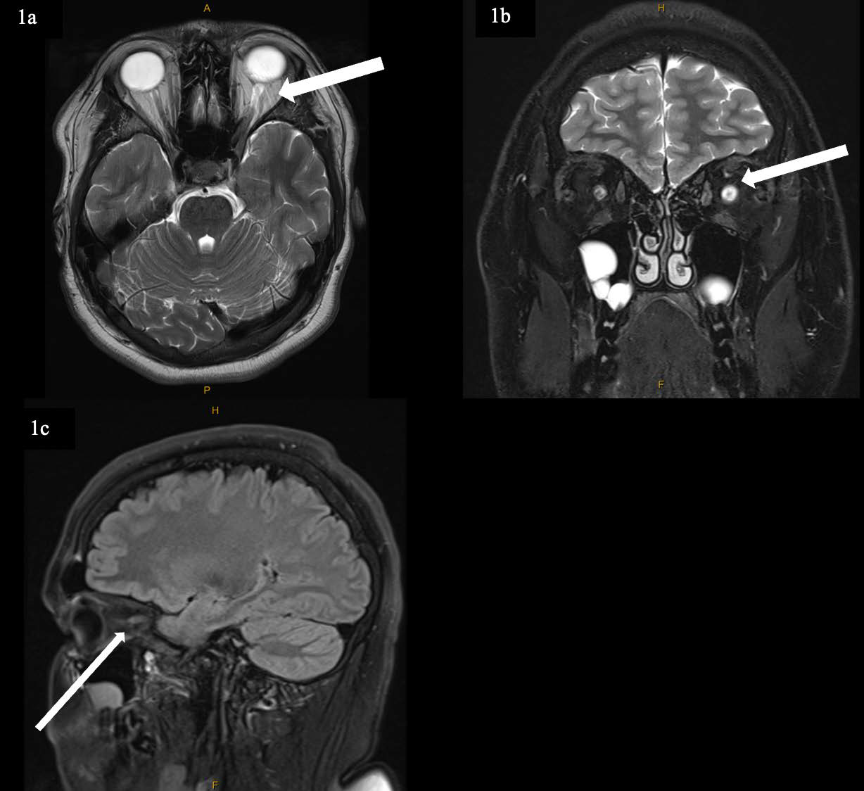
Case: A 19-year-old male presented with progressive, painless, unilateral (left) vision loss over one year. Initial MRI brain and orbits with gadolinium demonstrated increased T2 signal in the left optic nerve with the diagnostic impression being left optic neuritis. Initial laboratory testing excluded infectious and inflammatory aetiologies. Repeat MRI three months later demonstrated persistent enhancement of the left optic nerve. The possibility of optic nerve sheath meningioma was raised and therefore, a Ga-68 DOTATATE PET/CT was arranged. This demonstrated linear fusiform uptake (SUVmax 6.4) along the left optic nerve extending to the orbital apex, consistent with a somatostatin receptor expressing lesion such as an optic nerve sheath meningioma.
Conclusion: This case highlights the diagnostic value of Ga-68 DOTATATE PET/CT in confirming optic nerve sheath meningioma in young patients with atypical or equivocal MRI findings and negative inflammatory markers.
Downloads
Published
Issue
Section
License
Copyright (c) 2026 Journal of Radiology Case Reports

This work is licensed under a Creative Commons Attribution-NonCommercial-NoDerivatives 4.0 International License.
The publisher holds the copyright to the published articles and contents. However, the articles in this journal are open-access articles distributed under the terms of the Creative Commons Attribution-NonCommercial-NoDerivs 4.0 License, which permits reproduction and distribution, provided the original work is properly cited. The publisher and author have the right to use the text, images and other multimedia contents from the submitted work for further usage in affiliated programs. Commercial use and derivative works are not permitted, unless explicitly allowed by the publisher.


