
Calcific Tendinosis Mimicking Septic Arthritis and Myositis: Case Report and Literature Review
DOI:
https://doi.org/10.3941/jrcr.6092Abstract
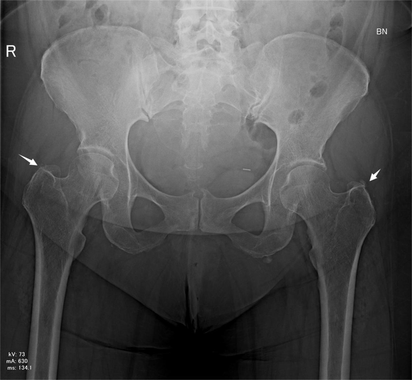
Hydroxyapatite deposition disease (HADD) is a crystal-induced condition that most commonly affects the shoulder, leading to rotator cuff tendinopathy. It rarely involves the hip joint and causes gluteus medius tendinitis, making diagnosis in such cases particularly challenging.
We report the case of a 54-year-old female patient who presented with acute, severe right hip pain and restricted mobility. Imaging and laboratory findings suggested right coxofemoral arthritis and focal myositis. Further evaluation revealed multifocal calcific tendinopathies, including a ruptured calcific tendinopathy of the gluteus medius tendon. Ultrasound-guided arthrocentesis ruled out septic arthritis.
The patient responded dramatically to colchicine, with rapid improvement in both pain and mobility. This case highlights the importance of considering HADD in the differential diagnosis of acute monoarthritis, particularly in atypical sites, and underscores the diagnostic value of bimodal imaging.
Downloads
Published
Issue
Section
License
Copyright (c) 2026 Journal of Radiology Case Reports

This work is licensed under a Creative Commons Attribution-NonCommercial-NoDerivatives 4.0 International License.
The publisher holds the copyright to the published articles and contents. However, the articles in this journal are open-access articles distributed under the terms of the Creative Commons Attribution-NonCommercial-NoDerivs 4.0 License, which permits reproduction and distribution, provided the original work is properly cited. The publisher and author have the right to use the text, images and other multimedia contents from the submitted work for further usage in affiliated programs. Commercial use and derivative works are not permitted, unless explicitly allowed by the publisher.


