MRI Features and Diagnostic Challenges in Abdominal Pregnancy and its Mimickers: A Case Series
DOI:
https://doi.org/10.3941/jrcr.6085Abstract
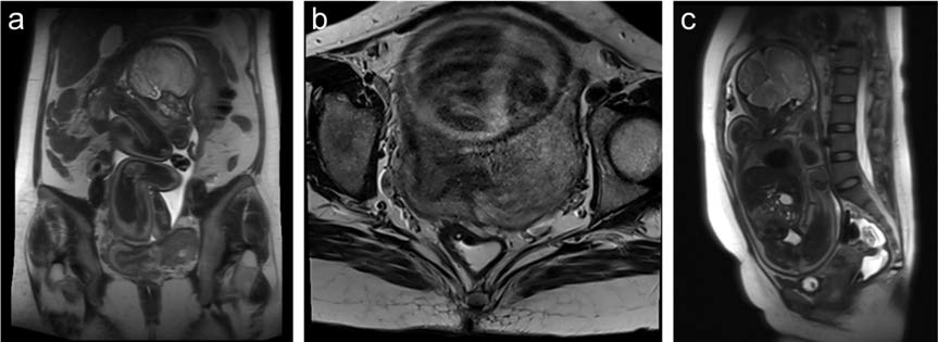
This study aims to present a case series of abdominal pregnancies and their mimickers, emphasizing the role of Magnetic Resonance Imaging (MRI) in diagnosis and management. The primary method used was evaluation with MRI for superior anatomical visualization. MRI successfully differentiated between an intrauterine pregnancy in a didelphic uterus (Case 1) and true abdominal pregnancies (Cases 2, 3, and 4). Key MRI features of abdominal pregnancy include: a thin-walled gestational sac, a placenta with irregular borders and heterogeneous texture, and vascularization originating from branches of the abdominal aorta. In conclusion, MRI is an invaluable imaging modality for diagnosing and assessing abdominal pregnancies. Diagnostic accuracy is enhanced by a systematic approach that includes careful anatomical assessment (aided by vaginal distension using hydrogel when necessary), analysis of placental and gestational sac wall characteristics, and evaluation of vascularization.
Downloads
Published
Issue
Section
License
Copyright (c) 2026 Journal of Radiology Case Reports

This work is licensed under a Creative Commons Attribution-NonCommercial-NoDerivatives 4.0 International License.
The publisher holds the copyright to the published articles and contents. However, the articles in this journal are open-access articles distributed under the terms of the Creative Commons Attribution-NonCommercial-NoDerivs 4.0 License, which permits reproduction and distribution, provided the original work is properly cited. The publisher and author have the right to use the text, images and other multimedia contents from the submitted work for further usage in affiliated programs. Commercial use and derivative works are not permitted, unless explicitly allowed by the publisher.