The Tumor Behind the Seizures: An Unexpected Diagnosis in Late Pregnancy
DOI:
https://doi.org/10.3941/jrcr.6054Abstract
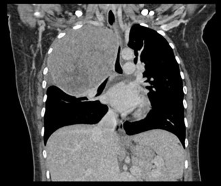
Schwannomas are benign nerve sheath tumors that rarely occur during pregnancy. We report the case of a 34-year-old woman at 36 weeks of gestation with gestational diabetes who presented with multiple seizures and was initially diagnosed with eclampsia. She also had dyspnea and a persistent cough. Chest radiography and contrast-enhanced computed tomography revealed a large right apical lung mass causing tracheal deviation. Magnetic resonance imaging showed a well-circumscribed heterogeneously enhancing lesion compressing the trachea, right main bronchus, and esophagus. Biopsy confirmed a schwannoma from the right T3 nerve root. She underwent resection via right thoracotomy, and histopathology verified the diagnosis. Despite successful surgery, she developed respiratory complications and died of pulmonary embolism. This rare case highlights the diagnostic challenge of schwannoma in pregnancy and the need to consider atypical causes of neurological and respiratory symptoms in such cases.
Downloads
Published
Issue
Section
License
Copyright (c) 2026 Journal of Radiology Case Reports

This work is licensed under a Creative Commons Attribution-NonCommercial-NoDerivatives 4.0 International License.
The publisher holds the copyright to the published articles and contents. However, the articles in this journal are open-access articles distributed under the terms of the Creative Commons Attribution-NonCommercial-NoDerivs 4.0 License, which permits reproduction and distribution, provided the original work is properly cited. The publisher and author have the right to use the text, images and other multimedia contents from the submitted work for further usage in affiliated programs. Commercial use and derivative works are not permitted, unless explicitly allowed by the publisher.