
A Different Kind of Marble: Sublingual Epidermoid Cyst with the "Sack of Marbles" Sign
DOI:
https://doi.org/10.3941/jrcr.6047Abstract
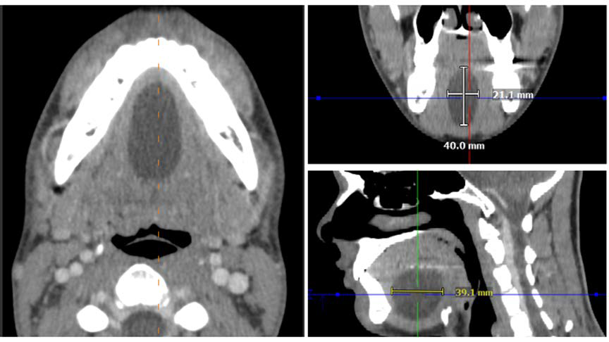
Epidermoid cysts account for approximately 1.6% of oral cavity cysts, with 7% occurring in the head and neck region. The “sack of marbles” appearance on magnetic resonance imaging has traditionally been considered specific for dermoid cysts, where fat globules from sebaceous secretions appear bright on T1-weighted sequences. A 22-year-old male presented with a midline floor-of-mouth mass discovered incidentally after trauma. The lesion enlarged over 18 months, causing mild speech difficulty. Imaging showed a well-circumscribed cyst with multiple bright foci and restricted diffusion, representing free-floating keratin globules. The appearance strongly suggested a dermoid cyst, but histopathology confirmed an epidermoid cyst without dermal appendages. This case demonstrates that epidermoid cysts can exhibit the “sack of marbles” pattern, highlighting that histopathologic correlation is essential for accurate diagnosis.
Downloads
Published
Issue
Section
License
Copyright (c) 2026 Journal of Radiology Case Reports

This work is licensed under a Creative Commons Attribution-NonCommercial-NoDerivatives 4.0 International License.
The publisher holds the copyright to the published articles and contents. However, the articles in this journal are open-access articles distributed under the terms of the Creative Commons Attribution-NonCommercial-NoDerivs 4.0 License, which permits reproduction and distribution, provided the original work is properly cited. The publisher and author have the right to use the text, images and other multimedia contents from the submitted work for further usage in affiliated programs. Commercial use and derivative works are not permitted, unless explicitly allowed by the publisher.


