
Minimally Invasive Management of Orbital Lymphangioma with Sclerotherapy: A Case Report
DOI:
https://doi.org/10.3941/jrcr.6030Abstract
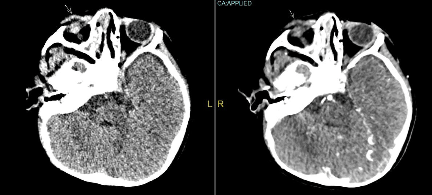
Orbital lymphangioma (OL) is a benign vascular disorder affecting the orbit and surrounding structures of the eye, typically appearing and progressing in childhood. Sclerotherapy has been reported to be the primary approach for OL, but with minimal evidenced reported in Indonesia. Therefore, this case report aims to present the case of a 3-year-old boy diagnosed with orbital lymphangioma who was treated with sclerotherapy. A 3-year-old male patient presented with progressive right eye enlargement over two months. The patient had a history of a congenital lump above the right ear, previously excised. Nutritional assessment showed age-appropriate growth with adequate dietary intake and no signs of malnutrition. The child attends pre-school with no concerns for peer bullying, and family/social support was good, with only intermittent school absence due to clinic visits. Examination showed right eye proptosis without visual disturbances or systemic symptoms. Imaging revealed a retro-orbital lymphangioma compressing orbital structures and signs of cerebral venous sinus thrombosis. Histopathology confirmed hemangio-lymphangioma. Sclerotherapy under general anesthesia was performed, followed by Trans Arterial Chemo Infusion (TACI) and Transcatheter Arterial Chemoembolization (TACE) evaluation. Post-treatment MRI showed reduced mass size and improvement in proptosis. Vision was preserved, recovery was uncomplicated, and functional/aesthetic outcomes improved, enabling return to normal daily activities and school participation. The patient continues under multidisciplinary care with favorable outcomes. There were functional and aesthetic improvements in a 3-year-old with orbital lymphangioma after sclerotherapy. Therefore, further research needs to focus on treatment protocols, molecular mechanisms, and long-term outcomes of sclerotherapy in lymphangioma orbitalis.
Downloads
Published
Issue
Section
License
Copyright (c) 2026 Journal of Radiology Case Reports

This work is licensed under a Creative Commons Attribution-NonCommercial-NoDerivatives 4.0 International License.
The publisher holds the copyright to the published articles and contents. However, the articles in this journal are open-access articles distributed under the terms of the Creative Commons Attribution-NonCommercial-NoDerivs 4.0 License, which permits reproduction and distribution, provided the original work is properly cited. The publisher and author have the right to use the text, images and other multimedia contents from the submitted work for further usage in affiliated programs. Commercial use and derivative works are not permitted, unless explicitly allowed by the publisher.


