
Utilizing Clinical Examination in Conjunction with Magnetic Resonance Cartilage Studies for Post-Operative Evaluation of Cartilage Repair: A Case Report
DOI:
https://doi.org/10.3941/jrcr.6008Abstract
Background
Articular cartilage is an essential component of our joints, and cartilage degeneration or injury may impede quality of life. With the advent of cartilage repair techniques first described in 1994, magnetic resonance imaging (MRI) has been the gold standard in evaluating cartilage repairs post-operatively. In particular, compositional MRI techniques such as T2 mapping are useful in qualitative and quantitative evaluation, due to its sensitivity for water and collagen content.
Case Report
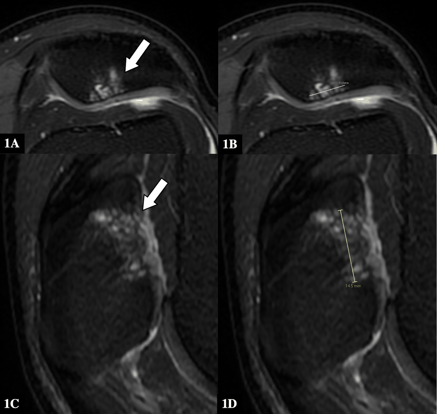
A 58-year-old male presented with left knee pain from a patella chondral defect at our tertiary institution and underwent a cartilage graft repair (HyaloFast; Anika Therapeutics, Italy). However, almost a year later, his left knee pain was noted to have recurred on follow-up. MRI cartilage studies showed features normally associated with graft viability, despite the patient’s persistent symptoms of knee pain on clinical examination.
He subsequently underwent a diagnostic arthroscopy, and the orthopedic surgeon noted that the surface of the previous cartilage graft was filled hyaline cartilage rather than fibrocartilage, and that there was no viable cartilaginous tissue at the site. The patient then had a revision cartilage patch repair surgery performed (ProChondrix CR; Stryker, USA). His left knee pain significantly improved after.
Notably, MRI cartilage T2RV mapping studies before and after the revision surgery were similar in appearance and showed features suggestive of graft viability, despite the clinical examination and history from the patient.
Conclusion
This case report serves to demonstrate how MRI T2RV mapping should not be solely utilized to assess technical success after cartilage repair surgery though it remains as a suitable non-invasive surveillance option. In addition to repeat MRI cartilage studies, a thorough history-taking to assess symptoms as well as physical examinations are critical in the post-operative follow-up.
Downloads
Published
Issue
Section
License
Copyright (c) 2026 Journal of Radiology Case Reports

This work is licensed under a Creative Commons Attribution-NonCommercial-NoDerivatives 4.0 International License.
The publisher holds the copyright to the published articles and contents. However, the articles in this journal are open-access articles distributed under the terms of the Creative Commons Attribution-NonCommercial-NoDerivs 4.0 License, which permits reproduction and distribution, provided the original work is properly cited. The publisher and author have the right to use the text, images and other multimedia contents from the submitted work for further usage in affiliated programs. Commercial use and derivative works are not permitted, unless explicitly allowed by the publisher.


