
Mesenchymal Chondrosarcoma Presenting as Deep Vein Thrombosis: A Case Report Highlighting Diagnostic Pitfalls and the Role of Imaging–Pathology Correlation
DOI:
https://doi.org/10.3941/jrcr.5995Abstract
Mesenchymal chondrosarcoma (MCS) is a rare, aggressive malignancy that can arise in either bone or soft tissue, typically affecting young adults in the second to third decades of life. Its hallmark biphasic histology—comprising undifferentiated small round cells interspersed with islands of cartilaginous differentiation—poses diagnostic challenges, as it can mimic other small round blue cell tumors. Because radiographic features are often nonspecific, accurate diagnosis requires careful integration of imaging, histopathologic, and immunohistochemical findings. We report the case of a healthy 26-year-old woman with a large juxtacortical mass arising from the left superior pubic ramus. Imaging revealed a lobulated, mineralized lesion with cortical origin and matrix calcification. This case is notable for its rare pelvic site and juxtacortical location, as well as its unusual initial presentation with iliofemoral deep venous thrombosis due to extrinsic venous compression. These findings underscore the diagnostic complexity of MCS and highlight the importance of considering this entity in the differential diagnosis of juxtacortical pelvic masses.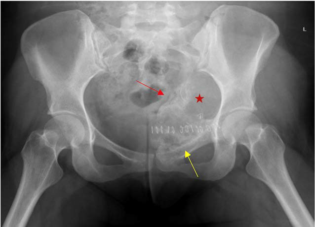
Downloads
Published
Issue
Section
License
Copyright (c) 2026 Journal of Radiology Case Reports

This work is licensed under a Creative Commons Attribution-NonCommercial-NoDerivatives 4.0 International License.
The publisher holds the copyright to the published articles and contents. However, the articles in this journal are open-access articles distributed under the terms of the Creative Commons Attribution-NonCommercial-NoDerivs 4.0 License, which permits reproduction and distribution, provided the original work is properly cited. The publisher and author have the right to use the text, images and other multimedia contents from the submitted work for further usage in affiliated programs. Commercial use and derivative works are not permitted, unless explicitly allowed by the publisher.


