
Lateral Sphenoidal Encephalocele as an Uncommon Cause of Trigeminal Neuralgia: A Case Report
DOI:
https://doi.org/10.3941/jrcr.5907Abstract
Introduction: Meningoencephaloceles are rare structural defects characterized by the herniation of brain tissue, meninges, and cerebrospinal fluid through bony defects in the skull base. While typically congenital, they may also result from trauma or chronically elevated intracranial pressure. Lateral sphenoidal encephaloceles, a rare subtype of basal encephaloceles, can present with neurological symptoms depending on their location. We describe a rare case of trigeminal neuralgia (TN) secondary to a lateral sphenoidal encephalocele, highlighting an underrecognized structural etiology of this condition.
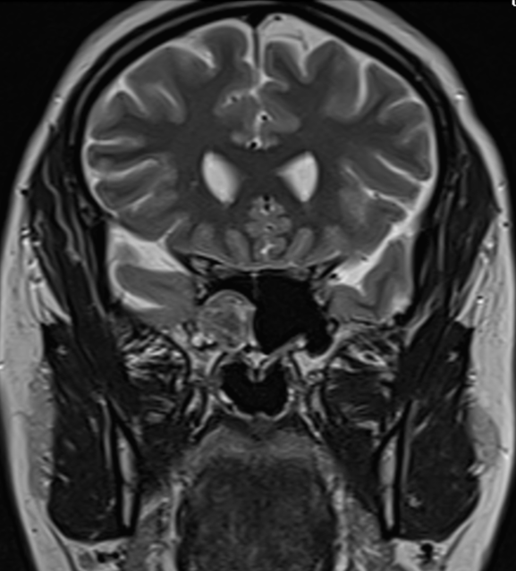
Case Presentation: A 35-year-old woman with a body mass index (BMI) of 37.4 presented with recurrent, paroxysmal electric shock-like pain localized to the proper trigeminal nerve distribution, consistent with classical TN. Initial brain MRI suggested a sphenoid sinus mucocele. However, a contrast-enhanced MRI performed one year later demonstrated herniation of gliotic temporal lobe tissue and enhancing meninges into the sphenoid sinus, consistent with a lateral sphenoidal encephalocele. CT imaging confirmed a bony defect in the lateral wall of the sphenoid sinus. The clinical and radiological findings supported a diagnosis of secondary TN due to the encephalocele. The patient was managed conservatively with oxcarbazepine, with partial symptom improvement.
Discussion: Lateral sphenoidal encephaloceles are rare but essential structural causes of trigeminal neuralgia, particularly due to their proximity to the trigeminal nerve. While classical TN is most often due to neurovascular compression, secondary causes such as encephaloceles should be considered in atypical cases. This case highlights the importance of contrast-enhanced imaging of the skull base in patients with unexplained facial pain, underscoring encephaloceles as a potentially underrecognized etiology.
Downloads
Published
Issue
Section
License
Copyright (c) 2026 Journal of Radiology Case Reports

This work is licensed under a Creative Commons Attribution-NonCommercial-NoDerivatives 4.0 International License.
The publisher holds the copyright to the published articles and contents. However, the articles in this journal are open-access articles distributed under the terms of the Creative Commons Attribution-NonCommercial-NoDerivs 4.0 License, which permits reproduction and distribution, provided the original work is properly cited. The publisher and author have the right to use the text, images and other multimedia contents from the submitted work for further usage in affiliated programs. Commercial use and derivative works are not permitted, unless explicitly allowed by the publisher.


