
Relapsed Primary Mediastinal Large B-Cell Lymphoma: The Crucial Role of PET-CT in Evaluating Disease Status and Treatment Response
DOI:
https://doi.org/10.3941/jrcr.5850Abstract
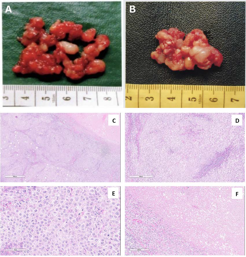
Despite advances in treating primary mediastinal large B-cell lymphoma, 20–30% of patients experience relapse or refractory disease. We report a case of a patient initially presenting with a neck lump, later diagnosed as primary mediastinal large B-cell lymphoma via biopsy. Initial positron emission tomography/computed tomography imaging showed a large fluorodeoxyglucose-avid anterior mediastinal mass. Following first-line chemotherapy, the disease was found to be refractory, requiring second-line treatment. The positron emission tomography/computed tomography played a key role in evaluating disease extent, treatment response, and relapse, indicated by new hypermetabolic nodes or extranodal lesions. It remains essential in guiding further therapy, including stem cell transplantation and brentuximab vedotin maintenance.
Downloads
Published
Issue
Section
License
Copyright (c) 2025 Journal of Radiology Case Reports

This work is licensed under a Creative Commons Attribution-NonCommercial-NoDerivatives 4.0 International License.
The publisher holds the copyright to the published articles and contents. However, the articles in this journal are open-access articles distributed under the terms of the Creative Commons Attribution-NonCommercial-NoDerivs 4.0 License, which permits reproduction and distribution, provided the original work is properly cited. The publisher and author have the right to use the text, images and other multimedia contents from the submitted work for further usage in affiliated programs. Commercial use and derivative works are not permitted, unless explicitly allowed by the publisher.


