
Imaging Features of Tube Misplacement in All Ages
DOI:
https://doi.org/10.3941/jrcr.5836Abstract
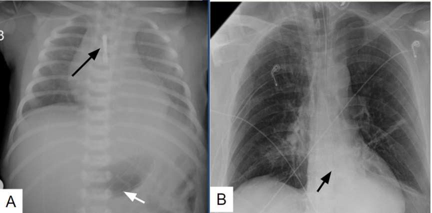
Although feeding tube misplacement is rare, its sequelae can be life threatening with significant morbidity and mortality. Increase in cases of gross feeding tube misplacement at our hospital in patients of all ages prompted investigation of misplacement rates and identification of risk factors. Literature review of enteric tube misplacement incidence and complications was performed. This exhibit illustrates the imaging spectrum of tube misplacement and complications both in the gastrointestinal tract and the tracheo-pulmonary tree. Common mimickers of tube misplacement are shown and differentiated. Risk factors for tube misplacement and how to prevent them are discussed at the end of this article.
Downloads
Published
Issue
Section
License
Copyright (c) 2025 Journal of Radiology Case Reports

This work is licensed under a Creative Commons Attribution-NonCommercial-NoDerivatives 4.0 International License.
The publisher holds the copyright to the published articles and contents. However, the articles in this journal are open-access articles distributed under the terms of the Creative Commons Attribution-NonCommercial-NoDerivs 4.0 License, which permits reproduction and distribution, provided the original work is properly cited. The publisher and author have the right to use the text, images and other multimedia contents from the submitted work for further usage in affiliated programs. Commercial use and derivative works are not permitted, unless explicitly allowed by the publisher.


