
What Can Imaging Do for Mycetoma in an Immunocompetent Patient in Temperate Region
DOI:
https://doi.org/10.3941/jrcr.5834Abstract
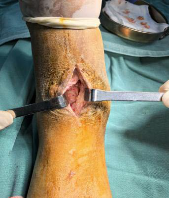
Mycetoma is a slowly persistent granulomatous infection of the cutaneous and deep subcutaneous tissue structures. It is caused by true fungi (eumycetoma) or bacteria (actinomycetoma), which is usually endemic in tropical countries. It may be misdiagnosed and delayed by low clinical suspicion, limited availability of diagnostic techniques, lack of biopsy and microbiological culture, potentially leading to disastrous consequences. Therefore, imaging plays a vital role in early recognition as a non-invasive technique, especially Magnetic resonance imaging, which demonstrates a hallmarked “dot-in-circle” sign of mycetoma. In this paper, we will present an immunocompetent patient with nearly two decades of history and review literature to highlight the importance of increasing awareness of mycetoma, particularly in non-endemic regions. The final diagnosis was made based on the characteristic “dot-in-circle” findings on Magnetic resonance images and pathologically confirmed.
Downloads
Published
Issue
Section
License
Copyright (c) 2025 Journal of Radiology Case Reports

This work is licensed under a Creative Commons Attribution-NonCommercial-NoDerivatives 4.0 International License.
The publisher holds the copyright to the published articles and contents. However, the articles in this journal are open-access articles distributed under the terms of the Creative Commons Attribution-NonCommercial-NoDerivs 4.0 License, which permits reproduction and distribution, provided the original work is properly cited. The publisher and author have the right to use the text, images and other multimedia contents from the submitted work for further usage in affiliated programs. Commercial use and derivative works are not permitted, unless explicitly allowed by the publisher.


