
Clinical Application of Echocardiography in Treatment of One Case of Mechanical Aortic Valve Stenosis with Giant Ventricular Aneurysm
DOI:
https://doi.org/10.3941/jrcr.5594Abstract
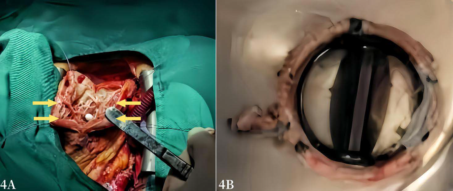
This paper reports a middle-aged woman who underwent mechanical aortic valve replacement surgery for congenital aortic valve malformation 7 years ago, and she had presented with exertional dyspnea and frequent ventricular arrhythmias in recent years. Transthoracic echocardiography revealed severe stenosis of the mechanical aortic valve orifice, as well as showed changes in left ventricular morphology caused by increased left ventricular afterload, which mainly manifested as localized hypertrophy of interventricular septum and a giant apical aneurysm. We used Simpson’s biplane method to pre-estimate the volume of left ventricular basal cavity, and assisted surgeons to make surgical plan in preliminarily.
During the operation, we assessed the shape, volume, and systolic function of left ventricular after left ventricular plasty, and determined that there were no potential risk factors for left ventricular outflow tract obstruction by transesophageal echocardiography. Under the guidance of transthoracic echocardiography, surgeons did not perform myocardial resection but performed mitral valve replacement. Finally, the patient was discharged smoothly.
Downloads
Published
Issue
Section
License
Copyright (c) 2026 Journal of Radiology Case Reports

This work is licensed under a Creative Commons Attribution-NonCommercial-NoDerivatives 4.0 International License.
The publisher holds the copyright to the published articles and contents. However, the articles in this journal are open-access articles distributed under the terms of the Creative Commons Attribution-NonCommercial-NoDerivs 4.0 License, which permits reproduction and distribution, provided the original work is properly cited. The publisher and author have the right to use the text, images and other multimedia contents from the submitted work for further usage in affiliated programs. Commercial use and derivative works are not permitted, unless explicitly allowed by the publisher.


