
Giant Pneumatized Ethmoid Bulla: A Rare Anatomical Variation
DOI:
https://doi.org/10.3941/jrcr.5563Abstract
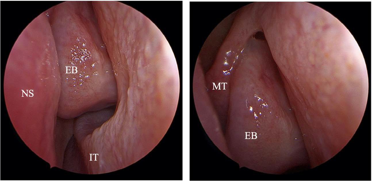
Pneumatization of the Ethmoid bulla varies greatly, and anatomical variations of the paranasal sinuses are quite common. However, giant ethmoid bulla prevalence in the published literature is relatively low. A 38-year-old woman presented with nasal obstruction and recurrent attacks of rhinosinusitis. The endoscopic examination showed a deviated nasal septum to the left, edematous left middle meatus, and hypertrophied inferior turbinates. The CT scan of the paranasal sinuses revealed an enlarged pneumatized ethmoid bulla, S-shaped nasal septum, bilateral inferior nasal turbinates hypertrophy, and obstructed left ostiomeatal complex (OMC), whereas the right OMC was patent. The patient underwent septoplasty and limited functional endoscopic sinus surgery. All symptoms were alleviated, and the patient remained symptom free after 6 months. Prospective studies should evaluate patients with sinonasal manifestations with radiologic evaluation complemented by nasal endoscopy to detect and rule out anatomical variations of the OMC region as a causative agent.
Downloads
Published
Issue
Section
License
Copyright (c) 2025 Journal of Radiology Case Reports

This work is licensed under a Creative Commons Attribution-NonCommercial-NoDerivatives 4.0 International License.
The publisher holds the copyright to the published articles and contents. However, the articles in this journal are open-access articles distributed under the terms of the Creative Commons Attribution-NonCommercial-NoDerivs 4.0 License, which permits reproduction and distribution, provided the original work is properly cited. The publisher and author have the right to use the text, images and other multimedia contents from the submitted work for further usage in affiliated programs. Commercial use and derivative works are not permitted, unless explicitly allowed by the publisher.


