
Assessing Benign Liver Lesions in Hepatitis B Patients: A Focus on Von Meyenburg Complexes
DOI:
https://doi.org/10.3941/jrcr.5558Abstract
Background: Von Meyenburg Complexes (VMCs), also known as biliary hamartomas, are rare, benign malformations of the intrahepatic bile ducts that can pose diagnostic challenges due to their resemblance to metastatic liver lesions, especially in patients with chronic hepatitis B virus (HBV) infection. Although generally asymptomatic, VMCs require careful imaging-based differentiation from malignant liver lesions, particularly in HBV patients at risk for hepatocellular carcinoma (HCC).
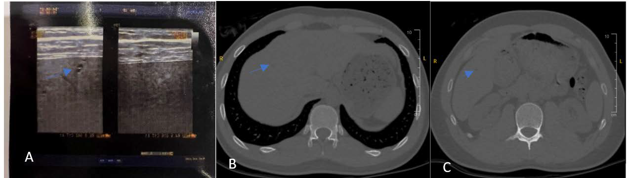
Case Presentation: We report the case of a 33-year-old male with chronic hepatitis B, normal liver function tests, and a viral load of 1.94 x 10^3 copies/mL. Screening ultrasound revealed a few subcentimeter liver cysts, prompting further evaluation. Non-contrast CT showed multiple hypoechoic structures scattered throughout the liver lobes, too small to characterize precisely. Magnetic resonance imaging (MRI) confirmed the presence of VMCs, displaying hypointensity on T1-weighted images and hyperintensity on T2-weighted images without enhancement, differentiating these lesions from malignancies. Three-dimensional MR cholangiography with maximum intensity projection showed additional, minute lesions with no involvement of the biliary tree, suggesting diffuse VMCs. Tumor markers were negative, further ruling out metastatic disease.
Discussion: This case emphasizes the importance of accurate imaging and diagnostic differentiation in patients with coexisting HBV and VMCs. VMCs have distinct MRI features that facilitate differentiation from malignant liver lesions, reducing the need for invasive biopsy. Given the patient’s hepatitis B status, a structured follow-up strategy was implemented, including routine ultrasound, elastography, and periodic liver function tests to monitor liver status without antiviral treatment.
Conclusion: The coexistence of VMCs and hepatitis B poses unique diagnostic challenges. Accurate imaging and a tailored follow-up approach are essential to ensure appropriate management and avoid unnecessary interventions. This case highlights the critical role of advanced MRI techniques in confirming benign lesions and guiding patient care in chronic liver disease contexts.
Downloads
Published
Issue
Section
License
Copyright (c) 2025 Journal of Radiology Case Reports

This work is licensed under a Creative Commons Attribution-NonCommercial-NoDerivatives 4.0 International License.
The publisher holds the copyright to the published articles and contents. However, the articles in this journal are open-access articles distributed under the terms of the Creative Commons Attribution-NonCommercial-NoDerivs 4.0 License, which permits reproduction and distribution, provided the original work is properly cited. The publisher and author have the right to use the text, images and other multimedia contents from the submitted work for further usage in affiliated programs. Commercial use and derivative works are not permitted, unless explicitly allowed by the publisher.


