
A Rare Case of Primary Extraosseous Osteosarcoma (EOS) of the thigh: A Case Report
DOI:
https://doi.org/10.3941/jrcr.5528Abstract
Background: Extraosseous osteosarcoma (EOO) is a rare mesenchymal malignancy, which produces osteoid, bone, or chondroid material and is located in the soft tissue without attachment to skeletal bones.
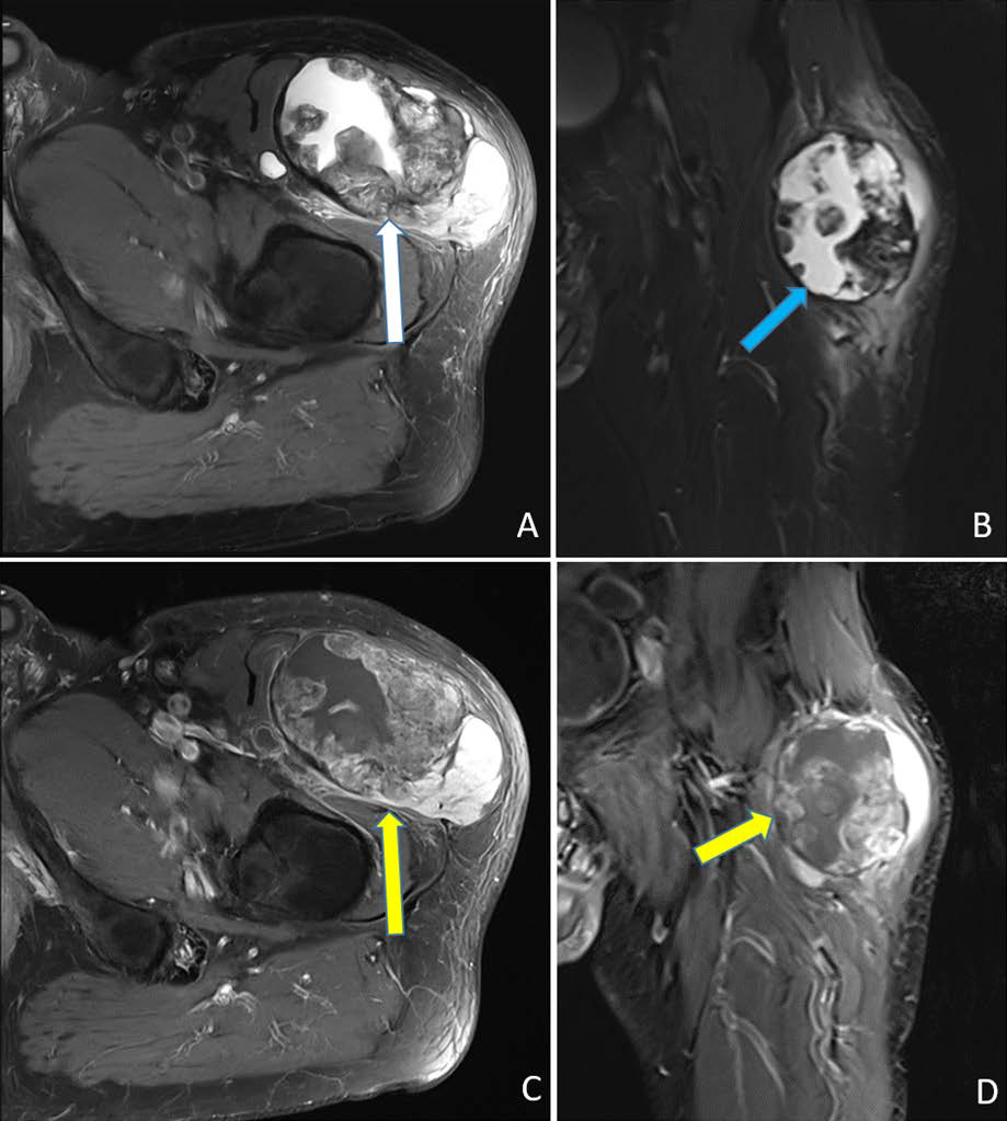
Case presentation: A 57-year-old male patient presented with extraosseous osteosarcoma located in the left rectus femoris muscle. The external magnetic resonance imaging revealed a large, irregular non-homogeneous contrast enhanced mass (largest diameter 9.5 cm). The final pathological diagnosis yielded extraosseous osteosarcoma. After interdisciplinary tumor board discussion, the following procedure was recommended: neoadjuvant systemic therapy with subsequent resection of the tumor and postoperative continuation of systemic therapy as well as discussion of adjuvant radiotherapy.
Conclusion: EOO should be treated as a soft tissue sarcoma with aggressive behavior and multimodality treatment should be actively sought to improve treatment outcome.
Downloads
Published
Issue
Section
License
Copyright (c) 2024 Journal of Radiology Case Reports

This work is licensed under a Creative Commons Attribution-NonCommercial-NoDerivatives 4.0 International License.
The publisher holds the copyright to the published articles and contents. However, the articles in this journal are open-access articles distributed under the terms of the Creative Commons Attribution-NonCommercial-NoDerivs 4.0 License, which permits reproduction and distribution, provided the original work is properly cited. The publisher and author have the right to use the text, images and other multimedia contents from the submitted work for further usage in affiliated programs. Commercial use and derivative works are not permitted, unless explicitly allowed by the publisher.


