MRI of Tailgut Cyst – Case Report and Review of Literature
DOI:
https://doi.org/10.3941/jrcr.5522Abstract
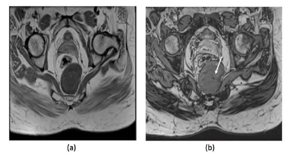
A tailgut cyst (TGC) is a rare congenital cystic developmental anomaly believed to arise from remnants of the embryonic tailgut. Most TGCs present as multicystic masses in the retrorectal space, with the potential for malignant transformation and spread into the ischioanal fossa. A significant number of cases are discovered incidentally during imaging. In contrast, others present with symptoms such as rectal bleeding, local mass effects on the rectum and bladder, abdominal pain, or constipation. Differential diagnoses include other cystic lesions such as dermoid cysts, epidermoid cysts, and sacrococcygeal teratomas. Diagnosis is primarily based on imaging, with MRI playing a critical role in distinguishing these lesions. Although numerous case reports describe imaging characteristics, very few emphasize the role of diffusion imaging in identifying tailgut cysts. Herein, we present a case of a tailgut cyst in a 43-year-old female, highlighting unique MRI features that aid in identifying the cyst and differentiating it from other cystic lesions.

Downloads
Published
Issue
Section
License
Copyright (c) 2024 Journal of Radiology Case Reports

This work is licensed under a Creative Commons Attribution-NonCommercial-NoDerivatives 4.0 International License.
The publisher holds the copyright to the published articles and contents. However, the articles in this journal are open-access articles distributed under the terms of the Creative Commons Attribution-NonCommercial-NoDerivs 4.0 License, which permits reproduction and distribution, provided the original work is properly cited. The publisher and author have the right to use the text, images and other multimedia contents from the submitted work for further usage in affiliated programs. Commercial use and derivative works are not permitted, unless explicitly allowed by the publisher.


