Pictorial Review of Unexpected and Extrathyroidal findings on Routine Thyroid Ultrasound Examinations
DOI:
https://doi.org/10.3941/jrcr.5505Abstract
Thyroid ultrasound is essential for the non-invasive assessment and management of thyroid diseases. While its primary focus is on evaluating the thyroid gland, incidental findings in the surrounding neck structures, known as extrathyroidal pathologies, are frequently encountered. These findings can have significant clinical implications and must not be overlooked.
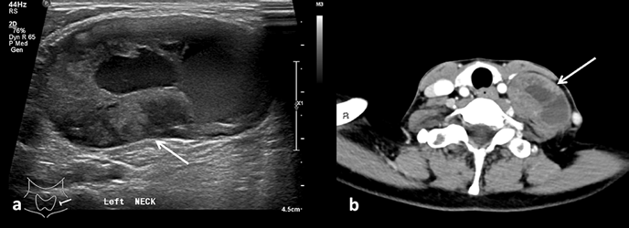
This retrospective review of 1,000 thyroid ultrasound examinations conducted between January 1, 2018 and December 31, 2023, aims to illustrate various extrathyroidal and unexpected findings. Notable examples include peripheral nerve sheath tumors (e.g., Schwannoma, plexiform neurofibromatosis), thyroglossal duct cysts, Zenker’s diverticulum, parathyroid adenomas, thyroid lymphomas, thrombus within the internal jugular vein, ectopic thymus and Zuckerkandl tubercle mimicking a thyroid mass.
While ultrasound remains the cornerstone imaging modality, in several cases, further evaluation with advanced imaging techniques such as computed tomography and magnetic resonance imaging was required, and some patients underwent surgical interventions.
The objective of this review is to highlight the importance of recognizing these unexpected findings during routine thyroid ultrasounds and to discuss their clinical management. Understanding the imaging characteristics of these pathologies enables radiologists to make accurate diagnoses, improving patient outcomes and reducing the need for unnecessary procedures.
This study underscores the role of multidisciplinary approaches in managing incidental extrathyroidal pathologies, emphasizing the combined use of imaging modalities to ensure effective and timely treatment planning.

Downloads
Published
Issue
Section
License
Copyright (c) 2024 Journal of Radiology Case Reports

This work is licensed under a Creative Commons Attribution-NonCommercial-NoDerivatives 4.0 International License.
The publisher holds the copyright to the published articles and contents. However, the articles in this journal are open-access articles distributed under the terms of the Creative Commons Attribution-NonCommercial-NoDerivs 4.0 License, which permits reproduction and distribution, provided the original work is properly cited. The publisher and author have the right to use the text, images and other multimedia contents from the submitted work for further usage in affiliated programs. Commercial use and derivative works are not permitted, unless explicitly allowed by the publisher.


