Schnitzler's Syndrome – An Uncommon Radiological Manifestation
DOI:
https://doi.org/10.3941/jrcr.5496Abstract
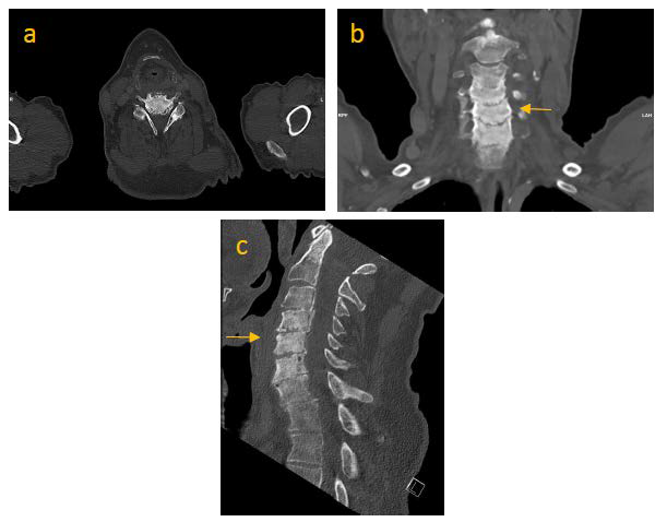
Schnitzler´s syndrome is a rare acquired autoinflammatory disorder. Chronic urticarial rash and monoclonal gammopathy (mainly IgM, rarely IgG [1]) are essential components of the disease. Schnitzler´s syndrome can present itself with osseous lesions, often in the distal femur and proximal tibia or in the hip bones. We report a case with uncommon radiological manifestation.

Downloads
Published
Issue
Section
License
Copyright (c) 2024 Journal of Radiology Case Reports

This work is licensed under a Creative Commons Attribution-NonCommercial-NoDerivatives 4.0 International License.
The publisher holds the copyright to the published articles and contents. However, the articles in this journal are open-access articles distributed under the terms of the Creative Commons Attribution-NonCommercial-NoDerivs 4.0 License, which permits reproduction and distribution, provided the original work is properly cited. The publisher and author have the right to use the text, images and other multimedia contents from the submitted work for further usage in affiliated programs. Commercial use and derivative works are not permitted, unless explicitly allowed by the publisher.


