Tenosynovial Giant Cell Tumor at the Upper Cervical Spine likely Arising from the Posterior Atlantoaxial Membrane: A Great Radiological Mimic
DOI:
https://doi.org/10.3941/jrcr.5463Abstract
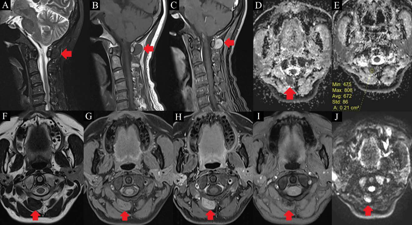
Although tenosynovial giant cell tumor (TSGCT) is commonly found in the limbs along tendon sheaths, bursae, and synovial joints, its occurrence in the axial skeleton is rare. Majority of the cases of TSGCT of the spine reported in the English literature were found to arise from the spinal facet joints. We report a rare case of TSGCT in the upper cervical spine likely arising from the posterior atlantoaxial membrane, complete with detailed magnetic resonance imaging (MRI) and computed tomography (CT) findings, and an approach to CT-guided biopsy of such paraspinal lesions.

Downloads
Published
Issue
Section
License
Copyright (c) 2024 Journal of Radiology Case Reports

This work is licensed under a Creative Commons Attribution-NonCommercial-NoDerivatives 4.0 International License.
The publisher holds the copyright to the published articles and contents. However, the articles in this journal are open-access articles distributed under the terms of the Creative Commons Attribution-NonCommercial-NoDerivs 4.0 License, which permits reproduction and distribution, provided the original work is properly cited. The publisher and author have the right to use the text, images and other multimedia contents from the submitted work for further usage in affiliated programs. Commercial use and derivative works are not permitted, unless explicitly allowed by the publisher.


