Mixed Atypical, Microcystic, And Angiomatous Meningioma: A Rare Radiologic And Histopathologic Findings
DOI:
https://doi.org/10.3941/jrcr.5431Abstract
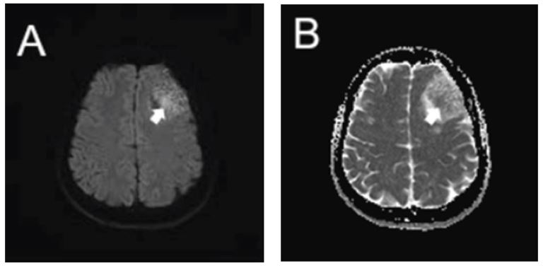
The occurrence of a mixed atypical, microcystic, and angiomatous meningioma is seldom encountered. To the best of the author's knowledge, no documented cases are reported to date that describe such a scenario. We report a 46-year-old female who presented to the emergency room after experiencing loss of consciousness preceded by a seizure and headache. Computed Tomography revealed a well-defined hypodense extra-axial mass in the left frontal region, described as an extra-axial cyst. Magnetic Resonance Imaging with intravenous contrast revealed a well-defined heterogeneous extra-axial lesion, isointense on T1-weighted images, hyperintense on T2-weighted images, and low intensity on Fluid Attenuated Inversion Recovery Images, consistent with the characteristics of a cyst. An irregular restricted diffusion area was observed in the center of the lesion on diffusion-weighted images. Striated vascular architecture was visible on the post-contrast T1 image. Histopathological findings of the tumor tissue confirm the diagnosis of mixed atypical, microcystic, and angiomatous meningioma. This case report discusses the radiological findings that provide diagnostic clues for meningiomas, specifically the atypical, microcystic, and angiomatous subtypes, along with their corresponding differential diagnoses.

Downloads
Published
Issue
Section
License
Copyright (c) 2024 Journal of Radiology Case Reports

This work is licensed under a Creative Commons Attribution-NonCommercial-NoDerivatives 4.0 International License.
The publisher holds the copyright to the published articles and contents. However, the articles in this journal are open-access articles distributed under the terms of the Creative Commons Attribution-NonCommercial-NoDerivs 4.0 License, which permits reproduction and distribution, provided the original work is properly cited. The publisher and author have the right to use the text, images and other multimedia contents from the submitted work for further usage in affiliated programs. Commercial use and derivative works are not permitted, unless explicitly allowed by the publisher.


