
Is it really a hematoma? Rare presentation of Invasive breast carcinoma
DOI:
https://doi.org/10.3941/jrcr.5406Abstract
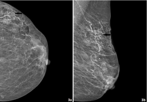
Breast cancer typically presents as a solid mass, architectural distortion, or calcifications on mammography, though it can also uncommonly present as a cystic mass based on subtype [1]. Hematoma usually presents as a cystic mass with internal echoes in hyperacute stage, subsequently turning to complicated cyst. In later stages, they appear as complex cysts with debris and thick echogenic wall with avascular mural nodule. Peripheral vascularity associated with the mass may represent interval inflammation. Clinical history of recent trauma or surgery is of great importance in the diagnosis. In the absence of recent history to account for the imaging findings, biopsy should be recommended [1]. This case report illustrates an unusual presentation of invasive ductal breast carcinoma as a hematoma in an elderly patient with history of frequent falls and offers several learning points to prevent misdiagnosis.
Downloads
Published
Issue
Section
License
Copyright (c) 2025 Journal of Radiology Case Reports

This work is licensed under a Creative Commons Attribution-NonCommercial-NoDerivatives 4.0 International License.
The publisher holds the copyright to the published articles and contents. However, the articles in this journal are open-access articles distributed under the terms of the Creative Commons Attribution-NonCommercial-NoDerivs 4.0 License, which permits reproduction and distribution, provided the original work is properly cited. The publisher and author have the right to use the text, images and other multimedia contents from the submitted work for further usage in affiliated programs. Commercial use and derivative works are not permitted, unless explicitly allowed by the publisher.


