A Case Report of Pericardial Inflammatory Myofibroblastic Tumor Involving the Inferior Vena Cava Causing Hepatomegaly
DOI:
https://doi.org/10.3941/jrcr.5396Abstract
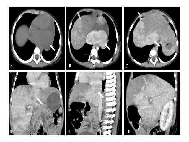
This case report discusses a 2-year-old child presenting with pericardial inflammatory myofibroblastic tumor (IMT) involving the inferior vena cava (IVC) causing hepatomegaly, which manifested as incidental upper abdominal distension without accompanying abdominal pain, nausea, or vomiting. Preoperative imaging revealed a soft tissue mass within the pericardium and pericardial effusion, causing deformation of the left atrium, and involving the IVC causing hepatomegaly. Subsequently, she underwent a mediastinal mass excisional biopsy by thoracotomy, which uncovered a smooth, resilient mass in the pericardium closely associated with the diaphragm. The tumor exhibited a fish-flesh appearance upon sectioning, with moderate pale red bloody fluid within the pericardium. The final pathology revealed an IMT. This case recapitulates the pathological classification, imaging findings, and differential diagnosis of the rare pericardial IMT

Downloads
Published
Issue
Section
License
Copyright (c) 2024 Journal of Radiology Case Reports

This work is licensed under a Creative Commons Attribution-NonCommercial-NoDerivatives 4.0 International License.
The publisher holds the copyright to the published articles and contents. However, the articles in this journal are open-access articles distributed under the terms of the Creative Commons Attribution-NonCommercial-NoDerivs 4.0 License, which permits reproduction and distribution, provided the original work is properly cited. The publisher and author have the right to use the text, images and other multimedia contents from the submitted work for further usage in affiliated programs. Commercial use and derivative works are not permitted, unless explicitly allowed by the publisher.


