
Incidental finding of delayed transformation of fibroadenoma to phyllodes tumor in young female: A Case Report
DOI:
https://doi.org/10.3941/jrcr.5385Abstract
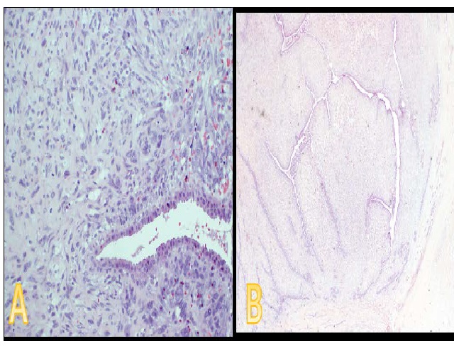
Phyllodes tumors arising from fibroadenomas are rare occurrences, particularly in young females. We present a case of a 27-year-old woman with a history of benign left breast fibroadenoma, who presented with right flank pain. Initial investigations revealed a significant enlargement of a previously identified benign left breast fibroadenoma. Imaging studies demonstrated a macrolobulated solid non-calcific lesion, notably larger than before, prompting referral to a breast clinic. Subsequent diagnostic ultrasound and contrast-enhanced MRI revealed a lesion displaying features concerning for phyllodes tumor. Biopsies performed in 2021 and at presentation (2022) indicated a transition from solitary benign fibroepithelial lesion (fibroadenoma) to borderline phyllodes tumor. Surgical excision lumpectomy was undertaken, with histological examination confirming borderline phyllodes tumor, stromal hyperplasia with mild cytologic atypia, and a notable mitotic rate of 9 per 10 high-power fields. This case underscores the importance of vigilance in monitoring fibroadenomas, as they may undergo transformation into phyllodes tumors.
Downloads
Published
Issue
Section
License
Copyright (c) 2024 Journal of Radiology Case Reports

This work is licensed under a Creative Commons Attribution-NonCommercial-NoDerivatives 4.0 International License.
The publisher holds the copyright to the published articles and contents. However, the articles in this journal are open-access articles distributed under the terms of the Creative Commons Attribution-NonCommercial-NoDerivs 4.0 License, which permits reproduction and distribution, provided the original work is properly cited. The publisher and author have the right to use the text, images and other multimedia contents from the submitted work for further usage in affiliated programs. Commercial use and derivative works are not permitted, unless explicitly allowed by the publisher.


