
Chondroblastoma-like Chondroma of the Temporomandibular Joint –Case Report with CT and MRI Findings
DOI:
https://doi.org/10.3941/jrcr.5366Abstract
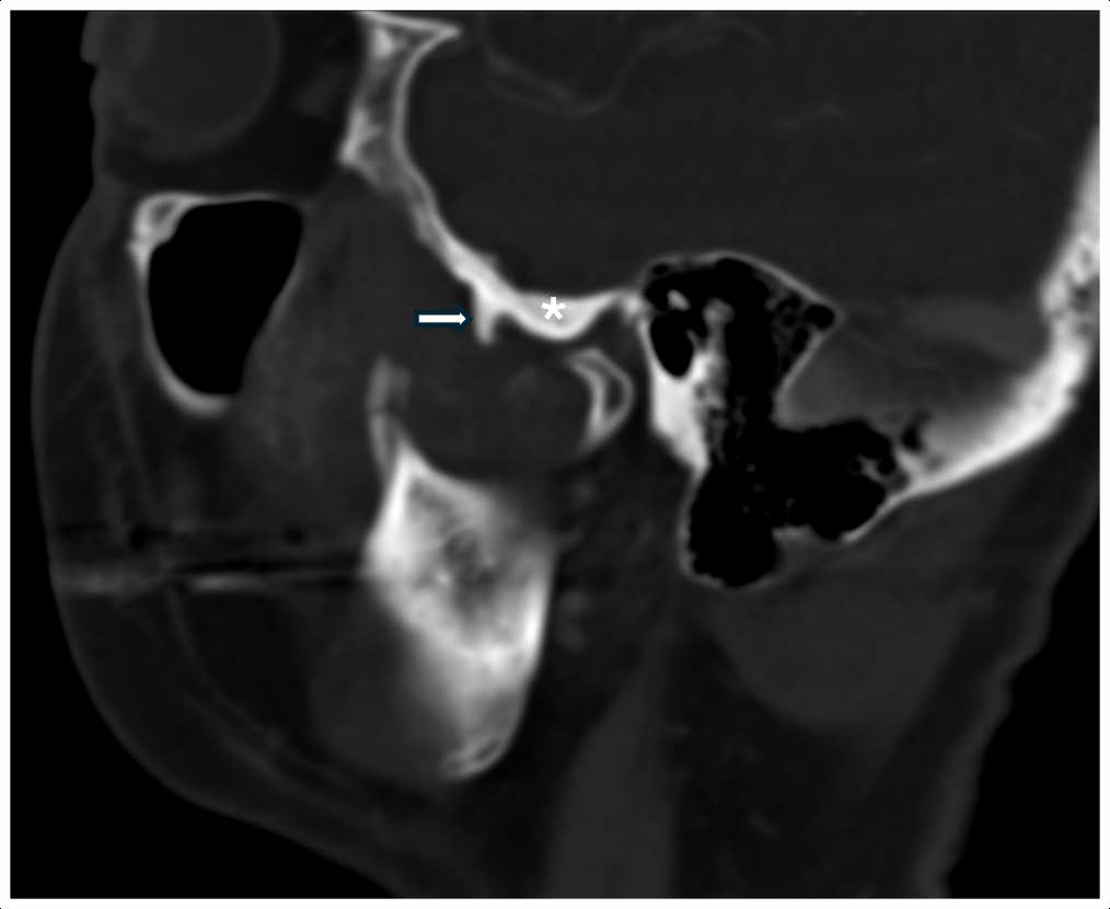
We present a case of a 58-year old man with a gradually enlarging right cheek mass. Computed tomography (CT) and magnetic resonance imaging (MRI) revealed a tumour with a chondroid matrix closely related to the temporomandibular joint. Subsequent surgical resection revealed a chondroblastoma-like chondroma. Identification of the the typical features of a chondroid matrix may aid in narrowing the differential diagnoses. Nonetheless, surgical resection is usually required to establish the definitive diagnosis.
Downloads
Published
Issue
Section
License
Copyright (c) 2024 Journal of Radiology Case Reports

This work is licensed under a Creative Commons Attribution-NonCommercial-NoDerivatives 4.0 International License.
The publisher holds the copyright to the published articles and contents. However, the articles in this journal are open-access articles distributed under the terms of the Creative Commons Attribution-NonCommercial-NoDerivs 4.0 License, which permits reproduction and distribution, provided the original work is properly cited. The publisher and author have the right to use the text, images and other multimedia contents from the submitted work for further usage in affiliated programs. Commercial use and derivative works are not permitted, unless explicitly allowed by the publisher.


