
Subcutaneous Forehead and Incidental Orbital Varices Diagnosed with MRI Under Sedation
DOI:
https://doi.org/10.3941/jrcr.v18i3.5353Abstract
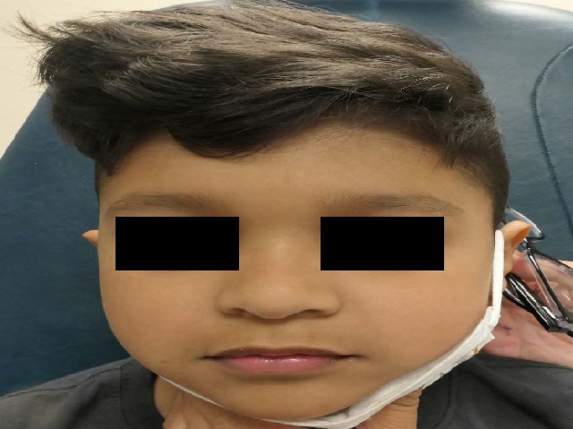
A 5-year-old boy presented for evaluation of an intermittent lesion in the left forehead that, per history from his mother, enlarged when the child slept on his right side. On clinical exam, although there were no notable findings with the child in the upright position, fullness and an enlarging mass when the child was placed on his right side could be reproduced. A diagnosis of a venous varix was suspected. Under sedation, a routine Magnetic Resonance Imaging of the orbits without and with intravenous contrast in the supine position and an axial T1 post-contrast in the right lateral decubitus position was obtained. The forehead lesion demonstrated a positional increase in size and had the Magnetic Resonance Imaging appearance of a varix. An incidental extraconal mass was also noted on Magnetic Resonance Imaging, which had the appearance of a varix and also increased in size in the right lateral decubitus position, confirming the diagnosis of an orbital varix. This case reviews the clinical presentation and imaging findings of two head and neck varices that were diagnosed with Magnetic Resonance Imaging under sedation using positional changes, thus, negating the need for radiation exposure to a child if Computed Tomography was utilized for the radiologic diagnosis.
Downloads
Published
Issue
Section
License
Copyright (c) 2024 Journal of Radiology Case Reports

This work is licensed under a Creative Commons Attribution-NonCommercial-NoDerivatives 4.0 International License.
The publisher holds the copyright to the published articles and contents. However, the articles in this journal are open-access articles distributed under the terms of the Creative Commons Attribution-NonCommercial-NoDerivs 4.0 License, which permits reproduction and distribution, provided the original work is properly cited. The publisher and author have the right to use the text, images and other multimedia contents from the submitted work for further usage in affiliated programs. Commercial use and derivative works are not permitted, unless explicitly allowed by the publisher.


