Parathyroid Carcinoma, an Uncommon Diagnosis
DOI:
https://doi.org/10.3941/jrcr.5339Abstract
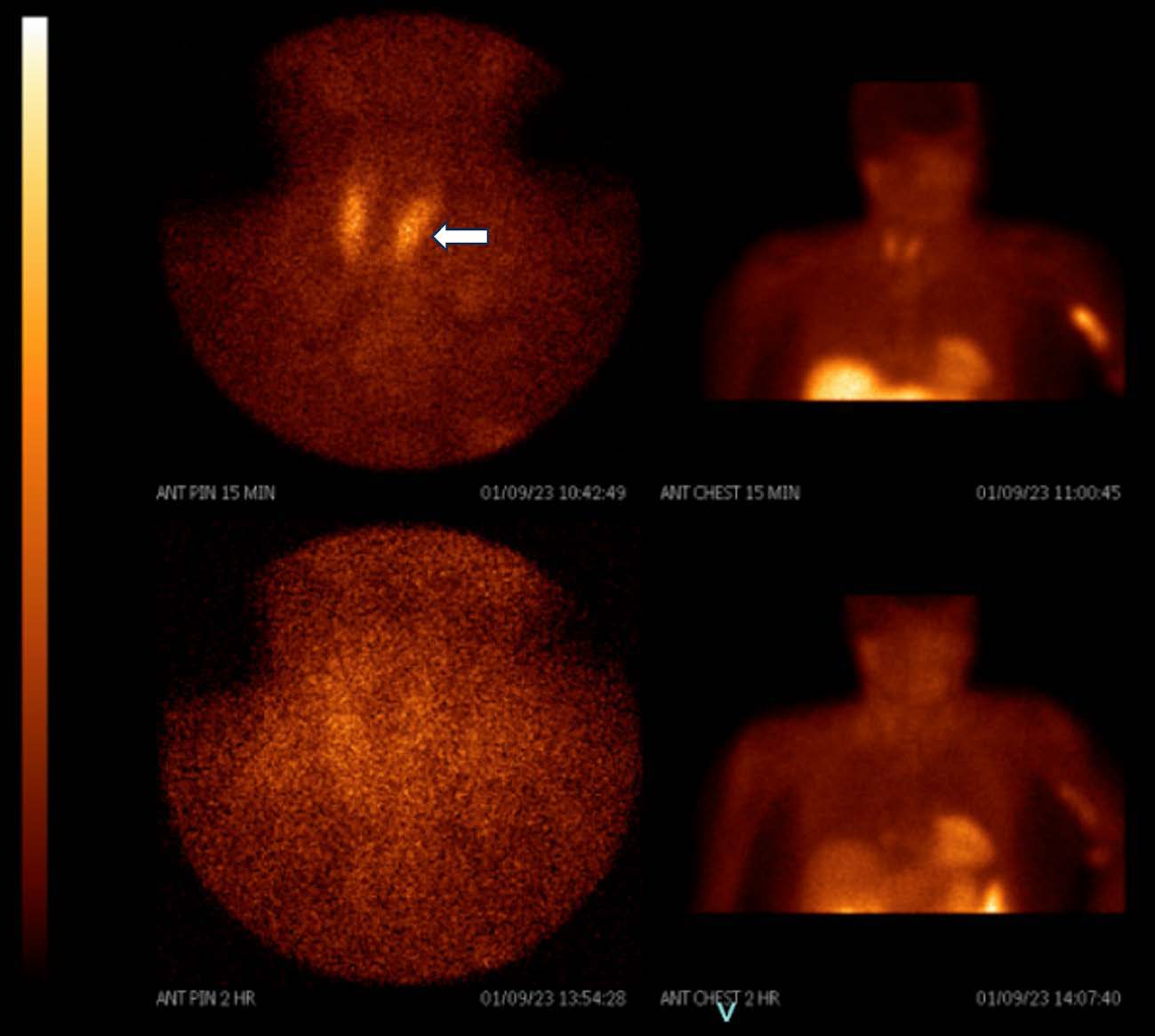
Parathyroid carcinoma should be considered in patients with clinical manifestations of severe hyperparathyroidism and hypercalcemia and corresponding abnormal laboratory values. Findings on several imaging modalities are highly suggestive of the diagnosis and often reveal malignant features, including local invasion or disseminated disease. Ultrasonography further aids in localizing the lesion and preoperative planning. Surgical resection, along with prospective histopathological examination, is the primary treatment. The roles of chemotherapy and radiation therapy remain controversial. Here, we present a case of a 67-year-old woman with histopathology-confirmed parathyroid carcinoma and multidisciplinary approach leading to diagnosis and management.

Downloads
Published
Issue
Section
License
Copyright (c) 2024 Journal of Radiology Case Reports

This work is licensed under a Creative Commons Attribution-NonCommercial-NoDerivatives 4.0 International License.
The publisher holds the copyright to the published articles and contents. However, the articles in this journal are open-access articles distributed under the terms of the Creative Commons Attribution-NonCommercial-NoDerivs 4.0 License, which permits reproduction and distribution, provided the original work is properly cited. The publisher and author have the right to use the text, images and other multimedia contents from the submitted work for further usage in affiliated programs. Commercial use and derivative works are not permitted, unless explicitly allowed by the publisher.


