
Biopsy Proven Cerebral Amyloid Related Inflammation Causing Vasogenic Edema with Midline Shift
DOI:
https://doi.org/10.3941/jrcr.5236Abstract
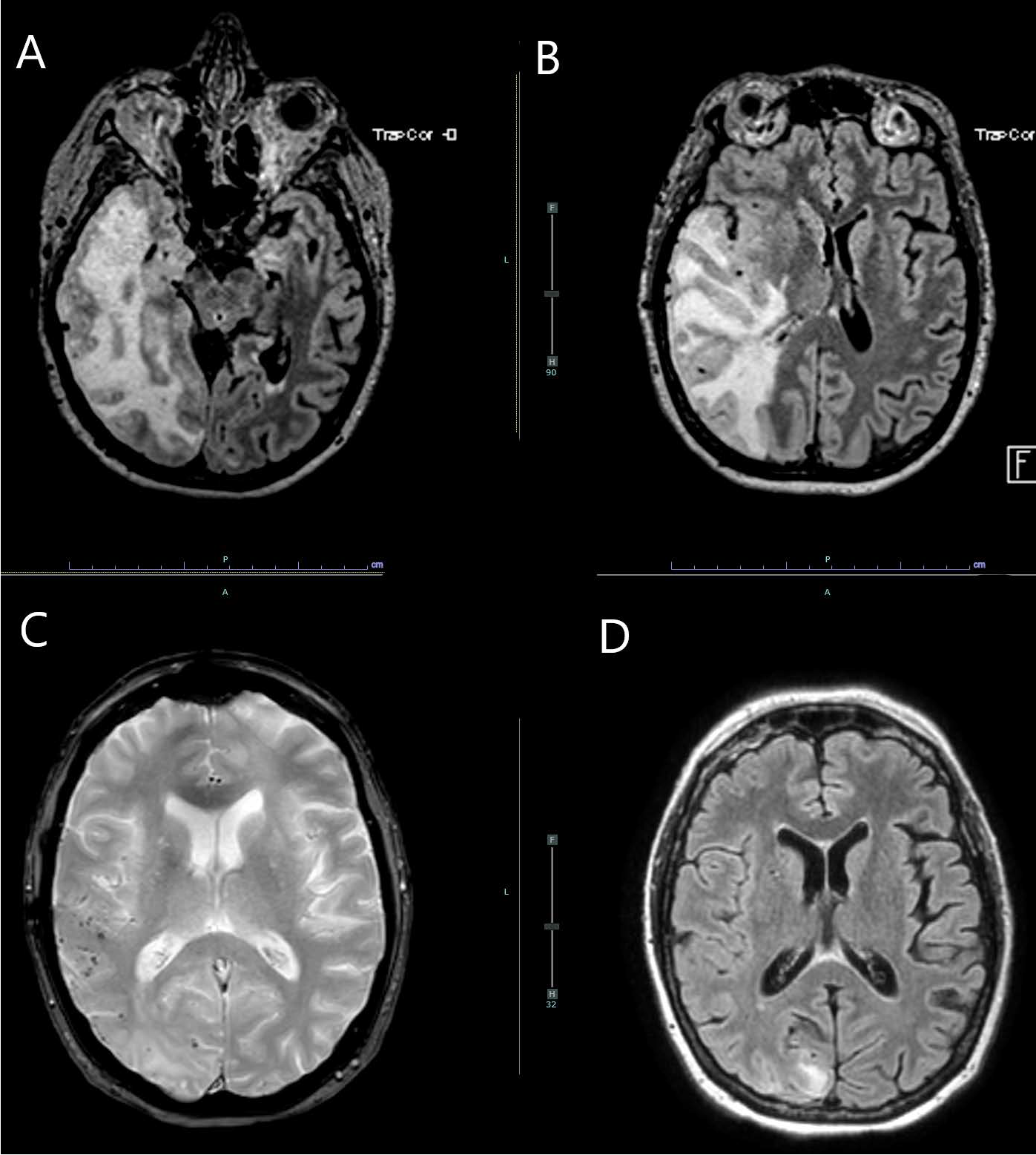
Cerebral amyloid angiopathy related inflammation is a rare condition that can present with rapid cognitive impairment, seizures, and headaches. Imaging typically shows the underling microbleeds and signs of inflammation; however, a brain biopsy is needed for a definitive diagnosis. Once diagnosed, treatment tends to be immunosuppression. Here, we present a severe case confirmed with brain biopsy with classic imaging of this infrequently seen condition.
Downloads
Published
Issue
Section
License
Copyright (c) 2026 Journal of Radiology Case Reports

This work is licensed under a Creative Commons Attribution-NonCommercial-NoDerivatives 4.0 International License.
The publisher holds the copyright to the published articles and contents. However, the articles in this journal are open-access articles distributed under the terms of the Creative Commons Attribution-NonCommercial-NoDerivs 4.0 License, which permits reproduction and distribution, provided the original work is properly cited. The publisher and author have the right to use the text, images and other multimedia contents from the submitted work for further usage in affiliated programs. Commercial use and derivative works are not permitted, unless explicitly allowed by the publisher.


