A Case Report of Uterine Torsion in a Postmenopausal Female with a Large Leiomyoma
DOI:
https://doi.org/10.3941/jrcr.v18i1.5035Keywords:
Uterine torsion, leiomyoma, gynecologic emergency, whirlpool sign, X-shaped, pelvic MRI, pelvic CTAbstract
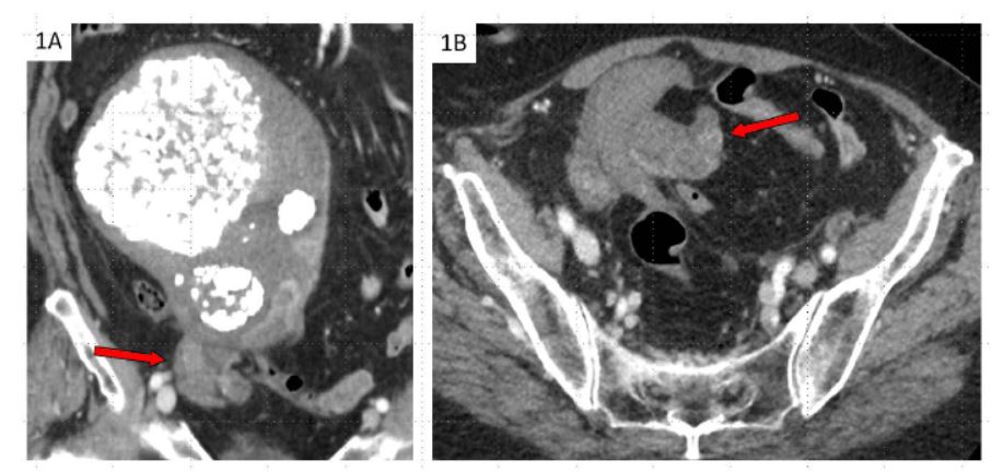
This case report discusses a diagnosis of uterine torsion in an 84-year-old woman who presented with five days of right lower quadrant abdominal pain, nausea, vomiting, constipation, and poor intake. Computed tomography (CT) imaging demonstrated a whorled configuration at the junction of the cervix and lower uterine segment, with the left gonadal vein crossing midline, and two previously known right leiomyomas now appearing on the left. These findings were consistent with the diagnosis of uterine torsion. She then underwent an urgent exploratory laparotomy, and the uterus was found to be dextroverted 270 degrees, with dark mottled purple tissue and engorged vessels. A supracervical hysterectomy and bilateral salpingo-oopherectomy were performed. Final pathology demonstrated extensive necrosis. This case reviews the classic presentation and imaging findings for the rare diagnosis of uterine torsion and options for management of both nongravid and gravid patients.

Downloads
Published
Issue
Section
License
Copyright (c) 2024 Journal of Radiology Case Reports

This work is licensed under a Creative Commons Attribution-NonCommercial-NoDerivatives 4.0 International License.
The publisher holds the copyright to the published articles and contents. However, the articles in this journal are open-access articles distributed under the terms of the Creative Commons Attribution-NonCommercial-NoDerivs 4.0 License, which permits reproduction and distribution, provided the original work is properly cited. The publisher and author have the right to use the text, images and other multimedia contents from the submitted work for further usage in affiliated programs. Commercial use and derivative works are not permitted, unless explicitly allowed by the publisher.


