
The Importance of Follow-Up: Juvenile Xanthogranuloma Mimicking Cephalohematoma
DOI:
https://doi.org/10.3941/jrcr.4829Keywords:
Juvenile Xanthogranuloma, Cephalohematoma, Follow-up care, UltrasonographyAbstract
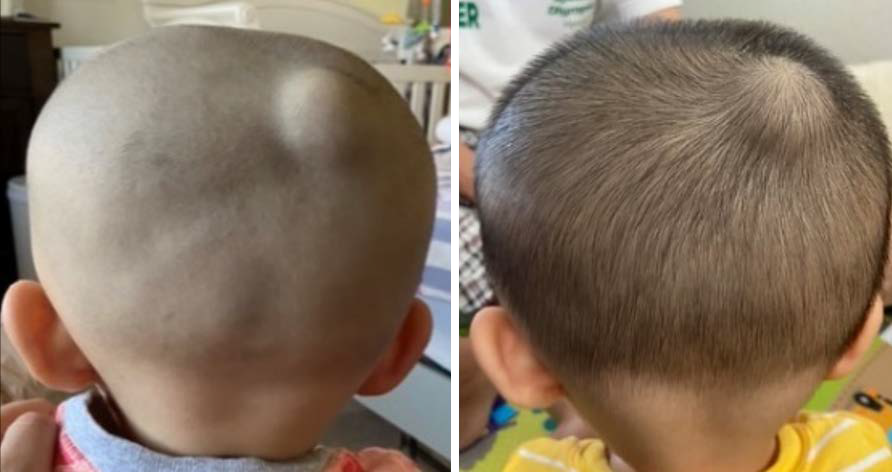
Juvenile xanthogranuloma is a type of non-Langerhans cell histiocytosis primarily affecting infants and young children. It is an uncommon disease and is rarely considered in the differential in radiological studies of infants. A case of juvenile xanthogranuloma mimicking a cephalohematoma in an otherwise healthy 3-month-old infant born via vacuum assisted delivery is presented here. At a follow-up appointment, interval growth and internal color Doppler flow with arterial waveform were noted on ultrasound. The differential diagnosis was expanded to include other well-circumscribed hypoechoic scalp lesions. At 8 months of age the lesion was surgically excised, and immunohistochemistry established the definitive diagnosis of juvenile xanthogranuloma. This case emphasizes the importance of following cephalohematomas clinically to resolution in order to exclude an alternate underlying pathology.
Downloads
Published
Issue
Section
License
Copyright (c) 2025 Journal of Radiology Case Reports

This work is licensed under a Creative Commons Attribution-NonCommercial-NoDerivatives 4.0 International License.
The publisher holds the copyright to the published articles and contents. However, the articles in this journal are open-access articles distributed under the terms of the Creative Commons Attribution-NonCommercial-NoDerivs 4.0 License, which permits reproduction and distribution, provided the original work is properly cited. The publisher and author have the right to use the text, images and other multimedia contents from the submitted work for further usage in affiliated programs. Commercial use and derivative works are not permitted, unless explicitly allowed by the publisher.


