
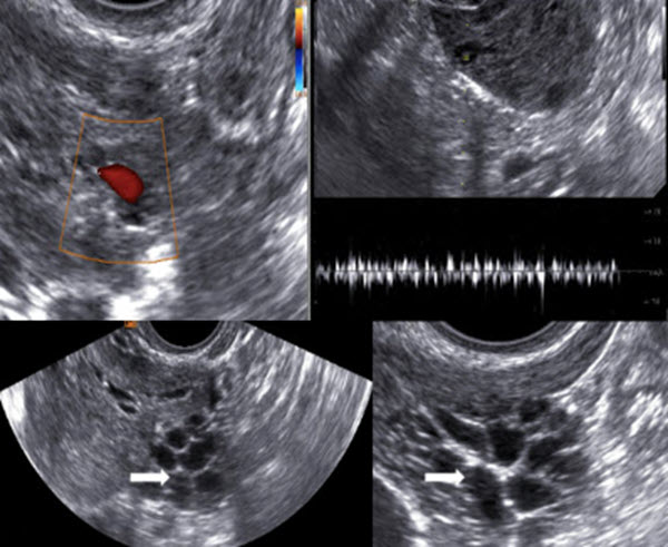
Real time ultrasound diagnosis of ovarian and pelvic filariasis by filarial dance sign
DOI:
https://doi.org/10.3941/jrcr.v17i7.4675Keywords:
ovarian filariasis, ultrasound, pelvis, filarial dance signAbstract
Parasitic infestations of the ovary are quite rare with ovary being the least common site of infection in the female genital tract. Filariasis is a parasitic disease caused by filarial nematodes (Wuchereria bancrofti, Brugia malayai, Brugia timori). It causes lymphatic obstruction with resultant edema and increase in the size of the affected organ. We report a case of 24-year-old married female who presented to our radiology department for ultrasound evaluation with the main aim being to look for retained products of conception after the termination of early pregnancy. However on ultrasound examination ovarian filariasis was an incidental diagnosis with the classical twirling movement (filarial dance sign) seen in one of the follicles of the ovary. Ultrasound is the imaging modality of choice for detecting the adult filarial worm/microfilaria in the lymphatic system. Ovarian filariasis is a very rare diagnosis with only a handful of cases being reported in literature with most cases being diagnosed incidentally on histopathological examination of the post operative specimen.
Downloads
Published
Issue
Section
License
Copyright (c) 2023 Journal of Radiology Case Reports

This work is licensed under a Creative Commons Attribution-NonCommercial-NoDerivatives 4.0 International License.
The publisher holds the copyright to the published articles and contents. However, the articles in this journal are open-access articles distributed under the terms of the Creative Commons Attribution-NonCommercial-NoDerivs 4.0 License, which permits reproduction and distribution, provided the original work is properly cited. The publisher and author have the right to use the text, images and other multimedia contents from the submitted work for further usage in affiliated programs. Commercial use and derivative works are not permitted, unless explicitly allowed by the publisher.


