
Venous Malformation in the Breast: Imaging Features to Avoid Unnecessary Biopsies or Surgery
DOI:
https://doi.org/10.3941/jrcr.v17i5.4635Keywords:
venous malformation, breast, mammography, light pressure ultrasonography, vascular disordersAbstract
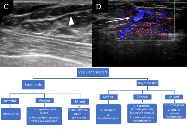
Venous malformations are now categorised under the broad heading of slow flow vascular malformations. They comprise abnormally dilated venous channels that fail to involute. These may be superficial or deep in location. We describe two cases of venous malformation in breast. Both the patients presented with focal pain in one breast. On mammography, they appeared as equal density well circumscribed soft tissue masses. No sonographic correlate was found on initial ultrasound examination. Subsequent ultrasonography performed by an experienced radiologist with minimal probe pressure revealed dilated veins. On the basis of imaging findings, the diagnosis of venous malformation was established.
Downloads
Published
Issue
Section
License
Copyright (c) 2023 Journal of Radiology Case Reports

This work is licensed under a Creative Commons Attribution-NonCommercial-NoDerivatives 4.0 International License.
The publisher holds the copyright to the published articles and contents. However, the articles in this journal are open-access articles distributed under the terms of the Creative Commons Attribution-NonCommercial-NoDerivs 4.0 License, which permits reproduction and distribution, provided the original work is properly cited. The publisher and author have the right to use the text, images and other multimedia contents from the submitted work for further usage in affiliated programs. Commercial use and derivative works are not permitted, unless explicitly allowed by the publisher.


