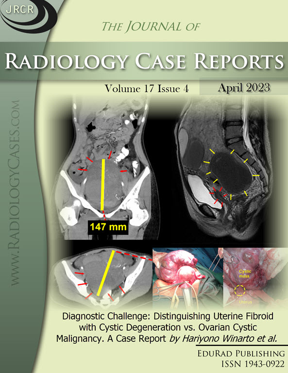
Diagnostic Challenge: Distinguishing Uterine Fibroid with Cystic Degeneration vs. Ovarian Cystic Malignancy. A Case Report
DOI:
https://doi.org/10.3941/jrcr.v17i4.4583Keywords:
Fibroid, Leiomyoma, Cystic Degeneration, Ovarian Cystic Malignancy, DiagnosisAbstract
Our case is a 24-year-old woman who has had abdominal enlargement for eleven months. She had an abdominal mass with an elevated level of CA-125 and imaging studies showed a pelvic cystic mass with a solid part, and thus malignancy was considered in the differential diagnosis. A laparotomy myomectomy was performed. Postoperative histopathological examination results revealed no signs of malignancy. In this case, both ultrasonography and magnetic resonance imaging could not visualize both ovaries and the stalk of the pedunculated fibroid on the posterior uterine corpus. On physical examination and imaging, cystic degeneration of uterine fibroid may present like an ovarian mass. Preoperative diagnosis is challenging. A definitive diagnosis is only feasible postoperatively following histological examination.Downloads
Published
2023-04-29
Issue
Section
Obstetric & Gynecologic Radiology
License
The publisher holds the copyright to the published articles and contents. However, the articles in this journal are open-access articles distributed under the terms of the Creative Commons Attribution-NonCommercial-NoDerivs 4.0 License, which permits reproduction and distribution, provided the original work is properly cited. The publisher and author have the right to use the text, images and other multimedia contents from the submitted work for further usage in affiliated programs. Commercial use and derivative works are not permitted, unless explicitly allowed by the publisher.


