
Isolated Spinal Cord Neurocysticercosis
DOI:
https://doi.org/10.3941/jrcr.v16i10.4543Keywords:
Neurocysticercosis, MRI, Spinal Cord, Brain, ParasiteAbstract
The incidence of neurocysticercosis is increasing in the US. The diagnosis is primarily made based on imaging findings, with clinical presentation and epidemiological exposure also playing a role. The differential diagnosis for neurocysticercosis (NCC) is extensive, and being able to differentiate between these conditions on imaging is crucial to making a proper diagnosis. Herein we present a case of a 37-year-old female who presented with lower extremity weakness and was found to have isolated spinal NCC. In this article, we will discuss the symptoms and imaging findings of neurocysticercosis to help guide diagnosis and management.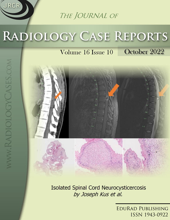
Downloads
Published
2022-10-31
Issue
Section
Neuroradiology
License
The publisher holds the copyright to the published articles and contents. However, the articles in this journal are open-access articles distributed under the terms of the Creative Commons Attribution-NonCommercial-NoDerivs 4.0 License, which permits reproduction and distribution, provided the original work is properly cited. The publisher and author have the right to use the text, images and other multimedia contents from the submitted work for further usage in affiliated programs. Commercial use and derivative works are not permitted, unless explicitly allowed by the publisher.


