
Male Breast Imaging Uncovers Lymphoma
DOI:
https://doi.org/10.3941/jrcr.v17i2.4508Keywords:
Lymphoma, Hodgkin, Breast, Mammography, MRI, Ultrasound, Lymphoproliferative, AxillaryAbstract
Background: A 36-year-old man presented with a palpable mass in the right axillary tail for four months. He was referred to breast imaging for diagnostic work-up. He does not have a family history of breast cancer. Aim: Breast imaging work-up for diagnosis of lymphoma is unusual and even more so in a male patient. Case presentation: After Breast Mammography and targeted Ultrasound of the axillary tail and axilla, Magnetic Resonance Imaging (MRI) was performed and suggested lymphoproliferative disorder. Excisional biopsy was performed after the breast MRI with removal of right axillary tissue measuring 15.0 x 5.5 x 2.0 cm and containing multiple lymph nodes. Excisional biopsy revealed Classic Hodgkin lymphoma of nodular sclerosis type. Staging [18F]-FDG PET/CT revealed early stage of disease. Conclusion: The presentation and diagnostic elements of Hodgkin Lymphoma are described in this case report emphasizing the significance of breast imaging in multiple populations.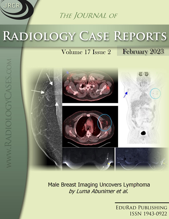
Downloads
Published
2023-02-28
Issue
Section
Breast Imaging
License
The publisher holds the copyright to the published articles and contents. However, the articles in this journal are open-access articles distributed under the terms of the Creative Commons Attribution-NonCommercial-NoDerivs 4.0 License, which permits reproduction and distribution, provided the original work is properly cited. The publisher and author have the right to use the text, images and other multimedia contents from the submitted work for further usage in affiliated programs. Commercial use and derivative works are not permitted, unless explicitly allowed by the publisher.


