
Imaging in hepatopulmonary syndrome- case report. A multicenter approach during the coronavirus pandemic
DOI:
https://doi.org/10.3941/jrcr.v17i10.4411Keywords:
Hepatopulmonary syndrome, macroaggregated albumin scan, liver transplantation, COVID-19, multi-centre approachAbstract
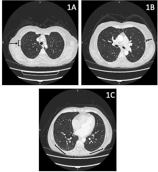
A 60-year-old lady with alcoholic liver disease developed central cyanosis and orthodeoxia. A technetium-99m macro-aggregated albumin lung perfusion scan and contrast echocardiogram were performed. A 13% right to left shunt was calculated from the macro-aggregated albumin scan. There were more bubbles in the left heart than the right at the end of the contrast echocardiogram. Hepatopulmonary syndrome was therefore diagnosed. The patient had a liver transplant five days after these investigations. Further discussion about hepatopulmonary syndrome will be provided.
Normally, macro-aggregated albumin scans are performed in few centers, however as this was at the height of the coronavirus pandemic, the scan needed to be performed locally to reduce the chance of the patient getting coronavirus. Local radiographers were remotely instructed on conducting the macro-aggregated albumin scan by a larger center to provide a timely and important investigation in a logistically difficult scenario.
Downloads
Published
Issue
Section
License
Copyright (c) 2023 Journal of Radiology Case Reports

This work is licensed under a Creative Commons Attribution-NonCommercial-NoDerivatives 4.0 International License.
The publisher holds the copyright to the published articles and contents. However, the articles in this journal are open-access articles distributed under the terms of the Creative Commons Attribution-NonCommercial-NoDerivs 4.0 License, which permits reproduction and distribution, provided the original work is properly cited. The publisher and author have the right to use the text, images and other multimedia contents from the submitted work for further usage in affiliated programs. Commercial use and derivative works are not permitted, unless explicitly allowed by the publisher.


