
In-utero Diagnosis of Double Encephalocele - Imaging Features and Review of Literature
DOI:
https://doi.org/10.3941/jrcr.v15i12.4230Keywords:
encephalocele, in-utero, MRI, fetal, cranium, double encephaloceleAbstract
Encephalocele is protrusion of brain parenchyma through a defect in the cranium. It is classified into various types based on the defect location: sincipital (fronto-ethmoidal), basal (trans-sphenoidal, spheno-ethmoidal, trans-ethmoidal, and spheno-orbital), occipital and parietal. Double encephaloceles are very rare with only a handful of cases reported in the literature and most of these cases involved either occipital or sub-occipital region. All, except one, cases of double encephaloceles were diagnosed postnatally. We present a case of double encephalocele with parietal and occipital components diagnosed in utero. To the best of our knowledge, this is the first case of double encephalocele involving the parietal and occipital skull bones diagnosed in-utero.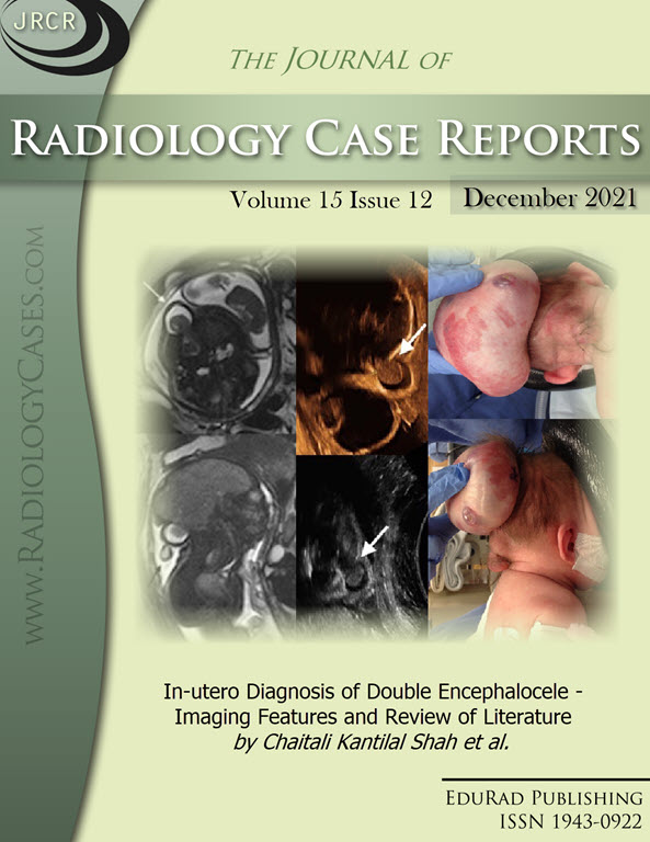
Downloads
Published
2021-12-31
Issue
Section
Neuroradiology
License
The publisher holds the copyright to the published articles and contents. However, the articles in this journal are open-access articles distributed under the terms of the Creative Commons Attribution-NonCommercial-NoDerivs 4.0 License, which permits reproduction and distribution, provided the original work is properly cited. The publisher and author have the right to use the text, images and other multimedia contents from the submitted work for further usage in affiliated programs. Commercial use and derivative works are not permitted, unless explicitly allowed by the publisher.


