
Implementation of 3D Printing in Medical Care for Preoperative Planning of Complex Ventricular Septal Defect
DOI:
https://doi.org/10.3941/jrcr.v15i11.4149Keywords:
Ventricular Septal Defect, 3D Modeling, 3D Printing, Presurgical planning, 3D Printing in Medicine, 3D Printing Applications in Cardiac ImagingAbstract
Three-dimensional (3D) modeling and printing in medicine have emerged to encompass every aspect of medical applications. This ranges from education, illustration, and treatment, as well as patient care whether for purposes of diagnosis or treatment and surgical planning. In the past few decades, these novel tools have shown promising utility to help radiologists and the medical team to improve quality of patient care and outcomes via 3D printing application and utilization. This workflow will be illustrated through a ventricular septal defect (VSD) case at which 3D analysis was critical in the assessment and treatment planning of the patient's underlying medical condition.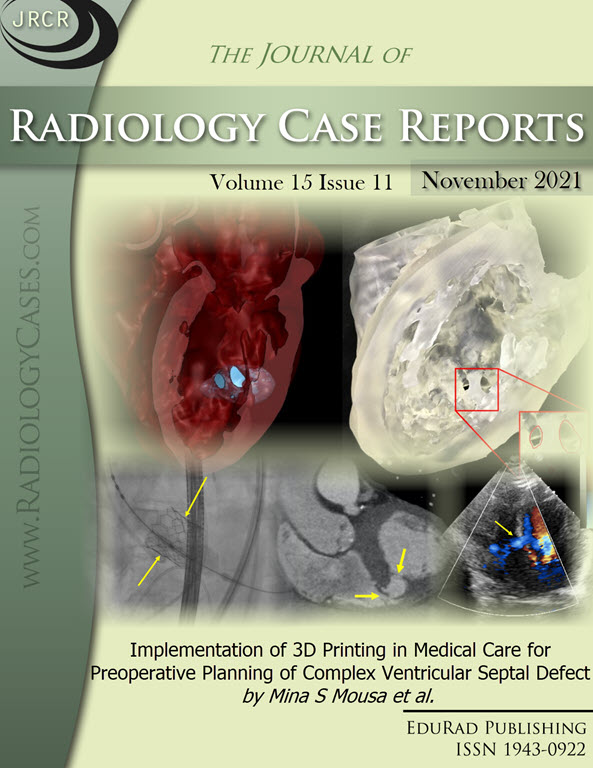
Downloads
Published
2021-11-30
Issue
Section
Cardiac Imaging
License
The publisher holds the copyright to the published articles and contents. However, the articles in this journal are open-access articles distributed under the terms of the Creative Commons Attribution-NonCommercial-NoDerivs 4.0 License, which permits reproduction and distribution, provided the original work is properly cited. The publisher and author have the right to use the text, images and other multimedia contents from the submitted work for further usage in affiliated programs. Commercial use and derivative works are not permitted, unless explicitly allowed by the publisher.


