
Diagnosis of Epipericardial Fat Necrosis on multimodality imaging in a pediatric patient: a case report and review of the literature
DOI:
https://doi.org/10.3941/jrcr.v14i5.3971Keywords:
Epipericardial fat necrosis, pericardium, pleuritic chest pain, Computed Tomography, Ultrasound, Magnetic Resonance Imaging, pediatricsAbstract
This is a case report of a 13-year-old male, presented to the Emergency Department complaining of a sudden onset left-sided pleuritic chest pain for 1 day. He was found to have a mass in the left Epipericardial fat with fat stranding and pleural effusion supporting the diagnosis of Epipericardial Fat Necrosis. The findings were established by Computed tomography and Ultrasound, and the final diagnosis was confirmed by Magnetic resonance imaging. Subsequently, the patient was discharged on analgesia; re-assessment one-month later showed clinical improvement with no symptom recurrence. Repeated Ultrasound demonstrated a marked decrease in size and echogenicity of the mass. In this paper we review the clinical and radiological manifestations of Epipericardial fat necrosis and the different management approaches taken over the years.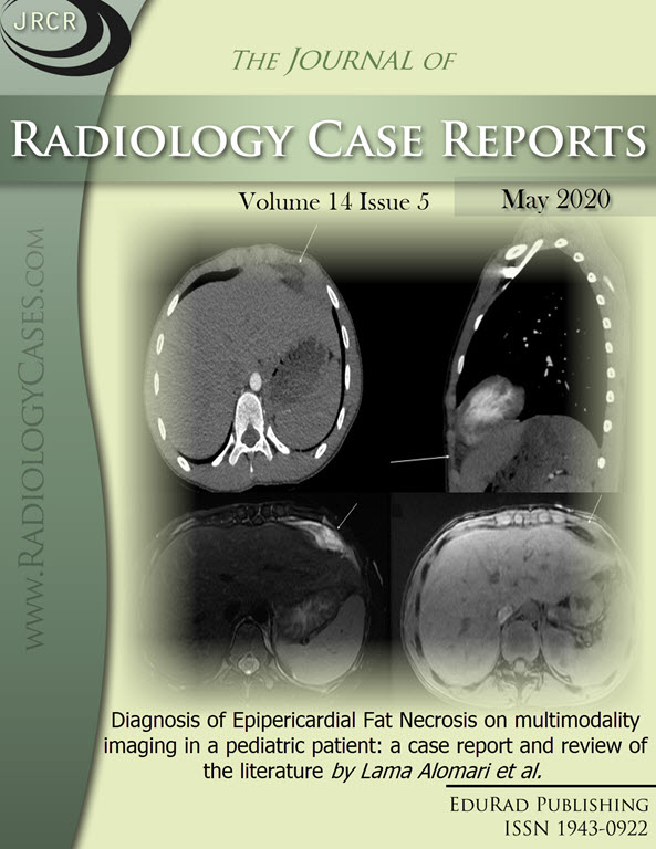
Downloads
Published
2020-05-26
Issue
Section
Pediatric Radiology
License
The publisher holds the copyright to the published articles and contents. However, the articles in this journal are open-access articles distributed under the terms of the Creative Commons Attribution-NonCommercial-NoDerivs 4.0 License, which permits reproduction and distribution, provided the original work is properly cited. The publisher and author have the right to use the text, images and other multimedia contents from the submitted work for further usage in affiliated programs. Commercial use and derivative works are not permitted, unless explicitly allowed by the publisher.


