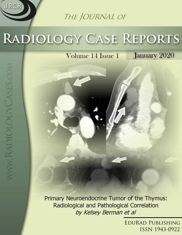
Primary Neuroendocrine Tumor of the Thymus: Radiological and Pathological Correlation
DOI:
https://doi.org/10.3941/jrcr.v14i1.3737Keywords:
Thymic neuroendocrine tumor, Thymic neoplasm, Thymus, Mediastinum, Magnetic resonance imaging, MRI, Computed tomography angiography, CTA, Positron emission tomography, PETAbstract
Primary neuroendocrine tumors of the thymus are extremely rare. In this report, we describe a case of a 69 year-old man with an intermediate grade thymic neuroendocrine tumor. The radiologic and histopathologic features of thymic neuroendocrine tumors are discussed with reference to relevant literature.Downloads
Published
2020-01-26
Issue
Section
Thoracic Radiology
License
The publisher holds the copyright to the published articles and contents. However, the articles in this journal are open-access articles distributed under the terms of the Creative Commons Attribution-NonCommercial-NoDerivs 4.0 License, which permits reproduction and distribution, provided the original work is properly cited. The publisher and author have the right to use the text, images and other multimedia contents from the submitted work for further usage in affiliated programs. Commercial use and derivative works are not permitted, unless explicitly allowed by the publisher.


