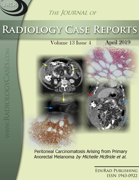
Peritoneal Carcinomatosis Arising from Primary Anorectal Melanoma
DOI:
https://doi.org/10.3941/jrcr.v13i4.3458Keywords:
Anorectal melanoma, anal melanoma, peritoneal carcinomatosis, ascites, peritoneum, omentum, Computed Tomography, Magnetic Resonance ImagingAbstract
Anorectal melanoma is a rare and aggressive malignancy with a poor prognosis. Anorectal melanoma makes up approximately 1 to 3% of all anorectal malignancies. There are no known risk factors for anorectal melanoma. Patients frequently experience a delay in diagnosis due to multiple factors including nonspecific symptoms and misdiagnosis for other benign entities. Anorectal melanoma has a high potential for distant metastases and radiographic imaging plays a key role in evaluating for metastatic disease. Common sites for metastasis include pelvic lymph nodes, lungs, liver, skin, and brain. We present a case report of a 75 year old female with a history of transanal excision of primary anorectal melanoma who presented with increasing abdominal pain and distention. Computed tomography scan of the abdomen and pelvis showed metastatic disease to the peritoneum with findings of extensive peritoneal carcinomatosis, demonstrating the aggressive nature of anorectal melanoma.Downloads
Published
2019-03-29
Issue
Section
Gastrointestinal Radiology
License
The publisher holds the copyright to the published articles and contents. However, the articles in this journal are open-access articles distributed under the terms of the Creative Commons Attribution-NonCommercial-NoDerivs 4.0 License, which permits reproduction and distribution, provided the original work is properly cited. The publisher and author have the right to use the text, images and other multimedia contents from the submitted work for further usage in affiliated programs. Commercial use and derivative works are not permitted, unless explicitly allowed by the publisher.


