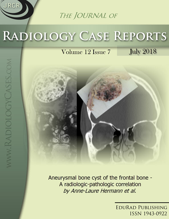
Aneurysmal bone cyst of the frontal bone - A radiologic-pathologic correlation
DOI:
https://doi.org/10.3941/jrcr.v12i7.3344Keywords:
Frontal aneurysmal bone cyst, ABC, CT and MRI findings, histopathologic correlation, USP6 rearrangementAbstract
We present a case of 27-year-old female who presented for a progressive frontal swelling with ipsilateral headache. Subsequent CT scan revealed an extradural and expansile multiloculated mass with thin and strongly enhanced septations and MRI evaluation showed internal hyperintensity on T2 with no restriction of diffusion and confirmed the multiple cystic spaces with enhancing septations and rare hemorrhagic fluid-fluid levels. Surgery was performed and diagnosis of aneurysmal bone cyst was made on frozen section. Identification of USP6 fusion gene by in situ hybridization technique permitted to confirm the diagnosis of primary ABC. Although aneurysmal bone cyst (ABC) of the skull is a very rare entity and accounts for 2-6% of all ABCs, we should think about it in front of osteolytic and cystic skull changes even with very few fluid-fluid levels. Following description of our case and differential diagnoses, we conduct a literature review of skull ABCs imaging characteristics and discuss the interest of USP6 rearrangement identification.Downloads
Published
2018-07-28
Issue
Section
Musculoskeletal Radiology
License
The publisher holds the copyright to the published articles and contents. However, the articles in this journal are open-access articles distributed under the terms of the Creative Commons Attribution-NonCommercial-NoDerivs 4.0 License, which permits reproduction and distribution, provided the original work is properly cited. The publisher and author have the right to use the text, images and other multimedia contents from the submitted work for further usage in affiliated programs. Commercial use and derivative works are not permitted, unless explicitly allowed by the publisher.


