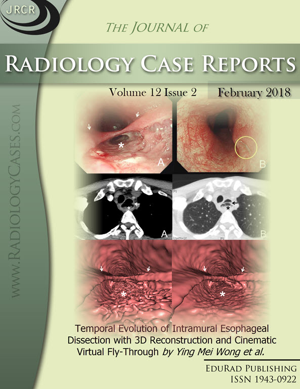
Temporal Evolution of Intramural Esophageal Dissection with 3D Reconstruction and Cinematic Virtual Fly-Through
DOI:
https://doi.org/10.3941/jrcr.v12i2.3288Keywords:
Computed Tomography, Conservative Treatment, Dissection, Esophageal Mucosa, HematomaAbstract
Intramural esophageal dissection is an uncommon condition, involving the separation of the esophageal mucosa from the muscular layers. To our knowledge, the temporal evolution of intramural esophageal dissection on computed tomography has not been previously demonstrated. We present a case of a 51-year-old male who first presented to the emergency department with fever, odynophagia, and dysphagia. He was treated for acute tonsillitis and discharged, but presented again after 10 days with worsening symptoms. A series of radiographs and computed tomography studies, with 3D reconstruction and cinematic virtual fly-through, in these 2 admissions depicts the temporal evolution of intramural hematoma to subsequent intramural esophageal dissection. Recognizing its appearance on imaging is invaluable in distinguishing it from other important differential diagnoses. A complete description of the case, relevant radiologic imaging, and review of the relevant literature are provided.Downloads
Published
2018-02-21
Issue
Section
Gastrointestinal Radiology
License
The publisher holds the copyright to the published articles and contents. However, the articles in this journal are open-access articles distributed under the terms of the Creative Commons Attribution-NonCommercial-NoDerivs 4.0 License, which permits reproduction and distribution, provided the original work is properly cited. The publisher and author have the right to use the text, images and other multimedia contents from the submitted work for further usage in affiliated programs. Commercial use and derivative works are not permitted, unless explicitly allowed by the publisher.


