
The Radiologic and Pathologic Diagnosis of Biphasic Pulmonary Blastoma
DOI:
https://doi.org/10.3941/jrcr.v11i9.3153Keywords:
Pulmonary Blastoma, Biphasic Pulmonary Blastoma, Pulmonary Neoplasm, Magnetic Resonance Imaging, Computed TomographyAbstract
Pulmonary blastomas are rare malignancies, representing 0.25% to 0.5% of all primary lung neoplasms with often aggressive progression and poor prognosis. Clinical management of pulmonary blastomas depends on histologic subtype, staging, and presentation, and may consist of surgery, chemotherapy, and radiation. Biphasic pulmonary blastoma is a subtype of pulmonary blastoma that exhibits biphasic histology, with both epithelial and mesenchymal malignant elements. We report a case of biphasic pulmonary blastoma in a 33-year-old female with 1 pack per day history of smoking for approximately 16 years, who presented with left-sided pleuritic chest pain on deep inspiration without otherwise significant pat medical history. Imaging evaluation using chest radiography, computed tomography, and magnetic resonance imaging identified a heterogenous, well-circumscribed, left lower lobe mass with extensive necrosis and hemorrhage. No lymphadenopathy or distant metastasis was detected through imaging evaluation. Surgical resection of the tumor followed by histopathological analysis confirmed a biphasic pulmonary blastoma.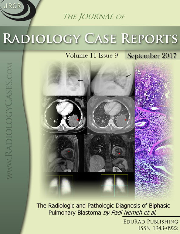
Downloads
Published
2017-09-25
Issue
Section
Thoracic Radiology
License
The publisher holds the copyright to the published articles and contents. However, the articles in this journal are open-access articles distributed under the terms of the Creative Commons Attribution-NonCommercial-NoDerivs 4.0 License, which permits reproduction and distribution, provided the original work is properly cited. The publisher and author have the right to use the text, images and other multimedia contents from the submitted work for further usage in affiliated programs. Commercial use and derivative works are not permitted, unless explicitly allowed by the publisher.


