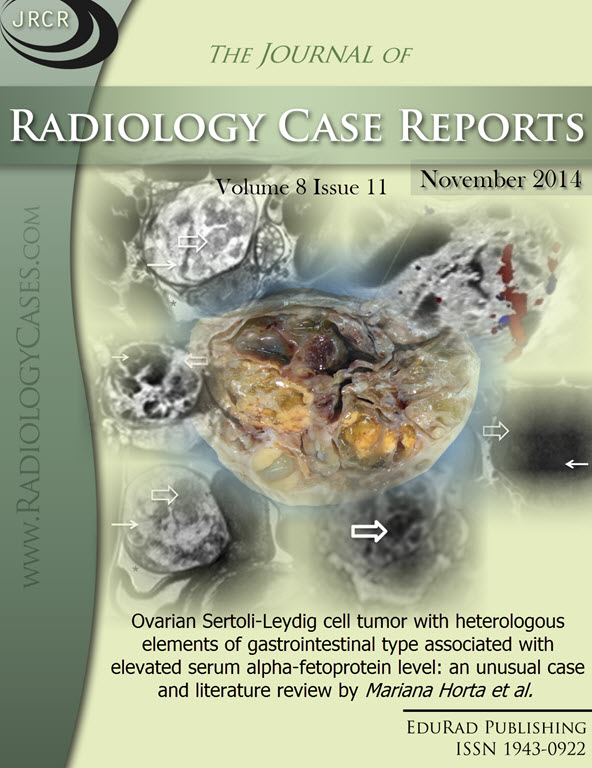
Ovarian Sertoli-Leydig cell tumor with heterologous elements of gastrointestinal type associated with elevated serum alpha-fetoprotein level: an unusual case and literature review
DOI:
https://doi.org/10.3941/jrcr.v8i11.2272Keywords:
Sertoli-Leydig tumor, Alpha-fetoprotein, Sex cord-stromal tumor, Ovarian tumor, Ultrasound, Magnetic resonanceAbstract
Here we describe the case of a 19-year-old woman with a poorly differentiated ovarian Sertoli-Leydig cell tumor and an elevated serum alpha-fetoprotein level. The patient presented with diffuse abdominal pain and bloating. Physical examination, ultrasound, and magnetic resonance imaging revealed a right ovarian tumor that was histopathologically diagnosed as a poorly differentiated Sertoli-Leydig cell tumor with heterologous elements. Her alpha-fetoprotein serum level was undetectable after tumor resection. Sertoli-Leydig cell tumors are rare sex cord-stromal tumors that account for 0.5% of all ovarian neoplasms. Sertoli-Leydig cell tumors tend to be unilateral and occur in women under 30 years of age. Although they are the most common virilizing tumor of the ovary, about 60% are endocrine-inactive tumors. Elevated serum levels of alpha-fetoprotein are rarely associated with Sertoli-Leydig cell tumors, with only approximately 30 such cases previously reported in the literature. The differential diagnosis should include common alpha-fetoprotein-producing ovarian entities such as germ cell tumors, as well as other non-germ cell tumors that have been rarely reported to produce this tumor marker.Downloads
Published
2014-11-24
Issue
Section
Obstetric & Gynecologic Radiology
License
The publisher holds the copyright to the published articles and contents. However, the articles in this journal are open-access articles distributed under the terms of the Creative Commons Attribution-NonCommercial-NoDerivs 4.0 License, which permits reproduction and distribution, provided the original work is properly cited. The publisher and author have the right to use the text, images and other multimedia contents from the submitted work for further usage in affiliated programs. Commercial use and derivative works are not permitted, unless explicitly allowed by the publisher.


