
Duplicated Pelvic Floor Musculature and Diastematomyelia in a Cloacal Exstrophy Patient
DOI:
https://doi.org/10.3941/jrcr.v8i10.2088Keywords:
Diastematomyelia, Spinal Dysraphism, Duplicated Pelvic Floor, Cloacal Exstrophy, Exstrophy-Epispadias Complex, EtiologyAbstract
Cloacal exstrophy is the most severe and rare form of the exstrophy-epispadias complex, presenting with exposed bladder halves extruding through an abdominal wall defect and variable genitourinary, gastrointestinal, musculoskeletal, and neurological defects. The authors report magnetic resonance imaging findings of a neurologically-intact, 24-month-old female with cloacal exstrophy who presented with anterior spinal dysraphism and diastematomyelia and duplicate pelvic floor musculature. The constellation of defects suggests a common genetic, biochemical, and embryological origin for duplication of the bladder, spinal cord, and pelvic floor muscles occurring in the fourth week of gestation.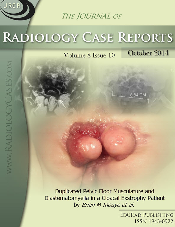
Downloads
Published
2014-10-19
Issue
Section
Pediatric Radiology
License
The publisher holds the copyright to the published articles and contents. However, the articles in this journal are open-access articles distributed under the terms of the Creative Commons Attribution-NonCommercial-NoDerivs 4.0 License, which permits reproduction and distribution, provided the original work is properly cited. The publisher and author have the right to use the text, images and other multimedia contents from the submitted work for further usage in affiliated programs. Commercial use and derivative works are not permitted, unless explicitly allowed by the publisher.


