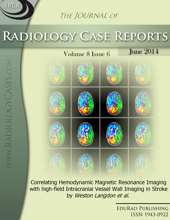
Correlating Hemodynamic Magnetic Resonance Imaging with high-field Intracranial Vessel Wall Imaging in Stroke
DOI:
https://doi.org/10.3941/jrcr.v8i6.1795Keywords:
Stroke, Cerebral Stroke, Cerebrovascular Accident, Cerebrovascular Stroke, Meningioma, Intracranial Meningioma, vessel-wall imaging, fMRI, Magnetic Resonance Imaging, Functional, MRI, AtherosclerosisAbstract
Vessel wall magnetic resonance imaging at ultra-high field (7 Tesla) can be used to visualize vascular lesions noninvasively and holds potential for improving stroke-risk assessment in patients with ischemic cerebrovascular disease. We present the first multi-modal comparison of such high-field vessel wall imaging with more conventional (i) 3 Tesla hemodynamic magnetic resonance imaging and (ii) digital subtraction angiography in a 69-year-old male with a left temporal ischemic infarct.Downloads
Published
2014-06-26
Issue
Section
Neuroradiology
License
The publisher holds the copyright to the published articles and contents. However, the articles in this journal are open-access articles distributed under the terms of the Creative Commons Attribution-NonCommercial-NoDerivs 4.0 License, which permits reproduction and distribution, provided the original work is properly cited. The publisher and author have the right to use the text, images and other multimedia contents from the submitted work for further usage in affiliated programs. Commercial use and derivative works are not permitted, unless explicitly allowed by the publisher.


