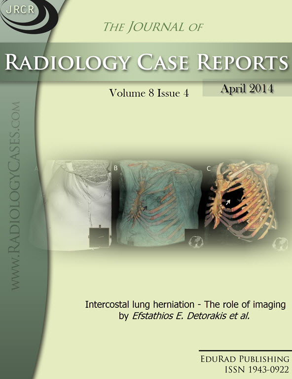
Intercostal lung herniation - The role of imaging
DOI:
https://doi.org/10.3941/jrcr.v8i4.1606Keywords:
Intercostal lung hernia, computed tomography, image reformatsAbstract
Extrathoracic lung hernias can be congenital or acquired. Acquired hernias may be classified by etiology into traumatic, spontaneous, and pathologic. We present a case of a 40-year-old male with a history of bronchial asthma and a blunt chest trauma who presented complaining of sharp chest pain of acute onset that began after five consecutive days of vigorous coughing. Upon physical examination a well-demarcated deformity overlying the third intercostal space of the left upper anterior hemithorax was revealed. Thoracic CT scan showed that a portion of the anterior bronchopulmonary segment of the left upper lobe had herniated through a chest wall defect. The role of imaging, especially chest computed tomography with multiplanar image reconstructions and maximum (MIP) and minimum intensity projection (MinIP) reformats can clearly confirm the presence of the herniated lung, the hernial sac, the hernial orifice in the chest wall, and exclude possible complications such as lung tissue strangulation.Downloads
Published
2014-04-26
Issue
Section
Thoracic Radiology
License
The publisher holds the copyright to the published articles and contents. However, the articles in this journal are open-access articles distributed under the terms of the Creative Commons Attribution-NonCommercial-NoDerivs 4.0 License, which permits reproduction and distribution, provided the original work is properly cited. The publisher and author have the right to use the text, images and other multimedia contents from the submitted work for further usage in affiliated programs. Commercial use and derivative works are not permitted, unless explicitly allowed by the publisher.


